. 1998 May;72(5):4095-103.
doi: 10.1128/JVI.72.5.4095-4103.1998.

A proline-rich motif (PPPY) in the Gag polyprotein of Mason-Pfizer monkey virus plays a maturation-independent role in virion release

Affiliations

A proline-rich motif (PPPY) in the Gag polyprotein of Mason-Pfizer monkey virus plays a maturation-independent role in virion release

J Yasuda et al. J Virol. 1998 May.

Abstract

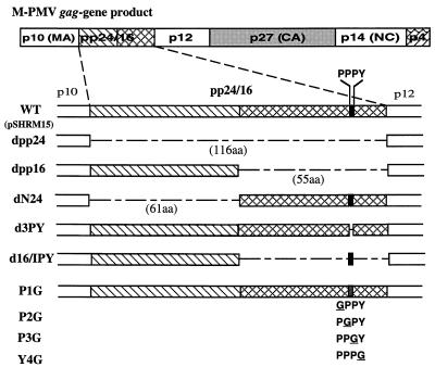
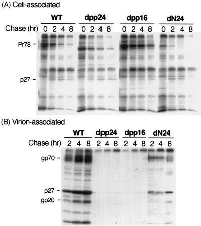
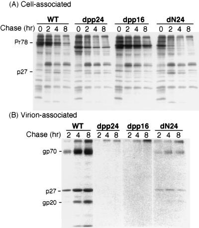
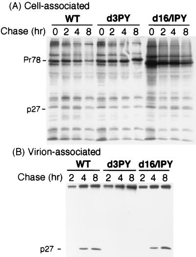
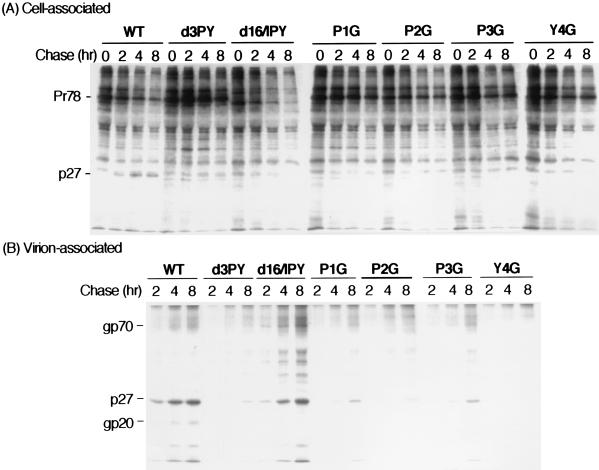
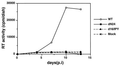
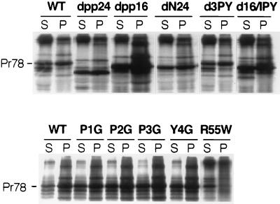
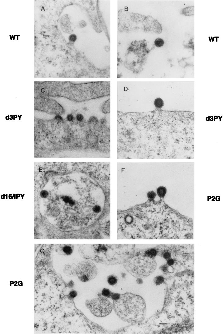
Virus assembly represents one of the last steps in the retrovirus life cycle. During this process, Gag polyproteins assemble at specific sites within the cell to form viral capsids and induce membrane extrusion (viral budding) either as assembly progresses (type C virus) or following formation of a complete capsid (type B and type D viruses). Finally, the membrane must undergo a fusion event to pinch off the particle in order to release a complete enveloped virion. Structural elements within the MA region of the Gag polyprotein define the route taken to the plasma membrane and direct the process of virus budding. Results presented here suggest that a distinct region of Gag is necessary for virus release. The pp24 and pp16 proteins of the type D retrovirus Mason-Pfizer monkey virus (M-PMV) are phosphoproteins that are encoded in the gag gene of the virus. The pp16 protein is a C-terminally located cleavage product of pp24 and contains a proline-rich motif (PPPY) that is conserved among the Gag proteins of a wide variety of retroviruses. By performing a functional analysis of this coding region with deletion mutants, we have shown that the pp16 protein is dispensable for capsid assembly but essential for virion release. Moreover, additional experiments indicated that the virus release function of pp16 was abolished by the deletion of only the PPPY motif and could be restored when this motif alone was reinserted into a Gag polyprotein lacking the entire pp16 domain. Single-amino-acid substitutions for any of the residues within this motif confer a similar virion release-defective phenotype. It is unlikely that the function of the proline-rich motif is simply to inhibit premature activation of protease, since the PPPY deletion blocked virion release in the context of a protease-defective provirus. These results demonstrate that in type D retroviruses a PPPY motif plays a key role in a late stage of virus budding that is independent of and occurs prior to virion maturation.


Figures

FIG. 1
FIG. 1
Representation of the pp24/16 deletion and the PPPY mutants. Dashed lines represent deleted regions. Filled boxes indicate the location of the PPPY motif. For the point mutants, substituted amino acids are underlined.
FIG. 2
FIG. 2
Pulse-chase analysis of pp24/16 deletion mutants in COS-1 cells. COS-1 cells were transfected with either WT (pSHRM15) or mutant DNAs and pulse-labeled with [3H]leucine for 30 min. After a 2-, 4-, or 8-h chase, virus-specific cell-associated (A) or virion-associated (B) proteins were immunoprecipitated with anti-Pr78 (Gag precursor) or anti-M-PMV antiserum, respectively. (A) In cells expressing either WT or mutant genomes, the Gag precursor polyproteins (Pr78) were labeled in the pulse (0 h) and the processed product (p27) appeared during chase periods. (B) Extracellular virions were pelleted from the culture fluids of chased cells. The positions of p27, the major capsid protein, and gp70 and gp20, the envelope glycoproteins, are shown.
FIG. 3
FIG. 3
Pulse-chase immunoprecipitation of HOS cells stably transfected with either WT or pp24/16 deletion mutant DNAs. Viral proteins from the cell lysates (A) and media (B) were analyzed as described in the legend to Fig. 2. Viral proteins were immunoprecipitated with anti-M-PMV antiserum. The migration positions of the WT viral proteins are indicated on the left.
FIG. 4
FIG. 4
Pulse-chase analysis of PPPY mutants in COS-1 cells. Viral proteins from the cell lysates (A) and media (B) were analyzed as described in the legend to Fig. 2, except anti-Pr78 antiserum was used for immunoprecipitation in both cases. The migration positions of the WT viral proteins are indicated on the left.
FIG. 5
FIG. 5
Pulse-chase analysis of PPPY point mutants in COS-1 cells. Viral proteins from the cell lysates (A) and media (B) were analyzed as described in the legend to Fig. 2. The migration positions of the WT viral proteins are indicated on the left.
FIG. 6
FIG. 6
Infectivity of mutant virions. Virus-containing culture medium from COS-1 cells transfected with either WT, dN24, or d16/IPY genomes was harvested and used to infect HOS cells. Infectivity was monitored as RT released over time following infection. Only WT showed any release of RT activity. In contrast, dN24 and d16/IPY showed no RT activity above that detected with the uninfected cells (Mock), demonstrating that these mutants are noninfectious.
FIG. 7
FIG. 7
Procapsid formation in WT- and mutant-transfected cells. COS-1 cells transfected with either WT or mutant DNA were pulse-labeled with [3H]leucine for 30 min and then chased in complete medium for 30 min. The cells were lysed, and assembled capsids were pelleted by centrifugation. Radiolabeled Gag polyproteins in the soluble (S) and pellet (P) fractions were immunoprecipitated with anti-Pr78 antiserum. WT Gag and all the mutant Gag precursors except R55W can be detected in both the soluble and pellet fractions.
FIG. 8
FIG. 8
Electron micrographs of COS-1 cells expressing WT and mutant viral genomes. Thin sections of COS-1 cells which had been transfected with WT or mutant DNA were examined under the electron microscope to determine the stage(s) at which mutant virus morphogenesis was blocked. (A and B) WT; (C and D) mutant d3PY; (E) d16/IPY; (F and G) P2G. Bar (panel G), 100 nm.
FIG. 9
FIG. 9
Pulse-chase analysis of a viral-protease-defective PPPY mutant in COS-1 cells. Viral proteins from the cell lysates (A) and media (B) were analyzed as described in the legend to Fig. 2. The migration positions of the WT viral proteins are indicated on the left.

Similar articles

Cited by

References

    1. Bedford M T, Chan D C, Leder P. FBP WW domains and the Abl SH3 domain bind to a specific class of proline-rich ligands. EMBO J. 1997;16:2376–2383. - PMC - PubMed
    1. Bork P, Sudol M. The WW domain: a signalling site in dystrophin? Trends Biochem Sci. 1994;19:531–533. - PubMed
    1. Bowles N, Bonnet D, Mulhauser F, Spahr P-F. Site-directed mutagenesis of the P2 region of the Rous sarcoma virus gag gene: effects on Gag polyprotein processing. Virology. 1994;203:20–28. - PubMed
    1. Bradac J, Hunter E. Polypeptides of Mason-Pfizer monkey virus. I. Synthesis and processing of the gag-gene products. Virology. 1984;138:260–275. - PubMed
    1. Brody B A, Rhee S S, Hunter E. Postassembly cleavage of a retroviral glycoprotein cytoplasmic domain removes a necessary incorporation signal and activates fusion activity. J Virol. 1994;68:4620–4627. - PMC - PubMed

Publication types

MeSH terms

LinkOut - more resources