. 1998 May;72(5):4192-204.
doi: 10.1128/JVI.72.5.4192-4204.1998.

The envelope protein encoded by the A33R gene is required for formation of actin-containing microvilli and efficient cell-to-cell spread of vaccinia virus

Affiliations

The envelope protein encoded by the A33R gene is required for formation of actin-containing microvilli and efficient cell-to-cell spread of vaccinia virus

R L Roper et al. J Virol. 1998 May.

Abstract

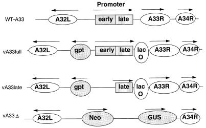
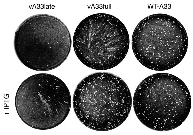
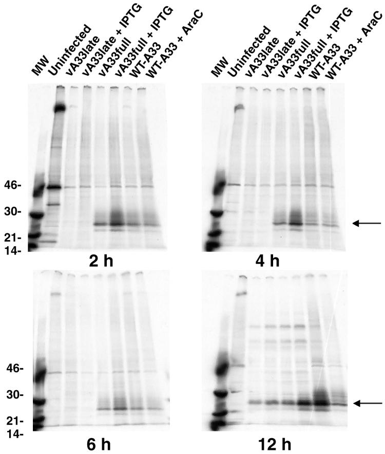
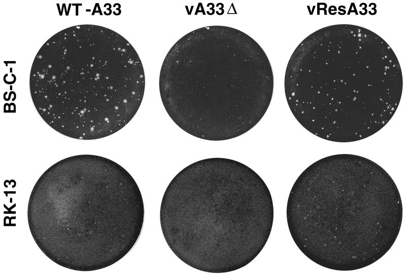
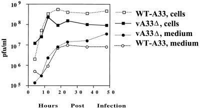
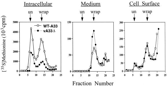
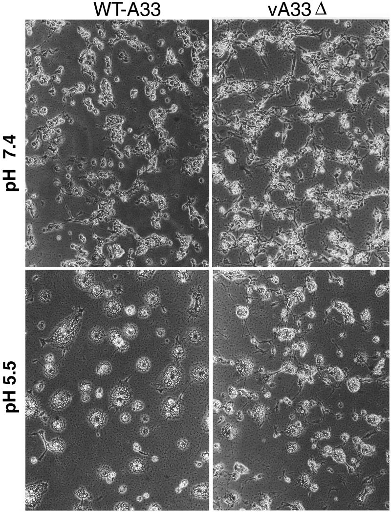
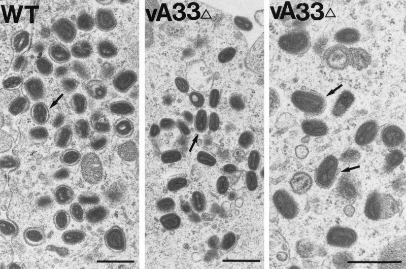
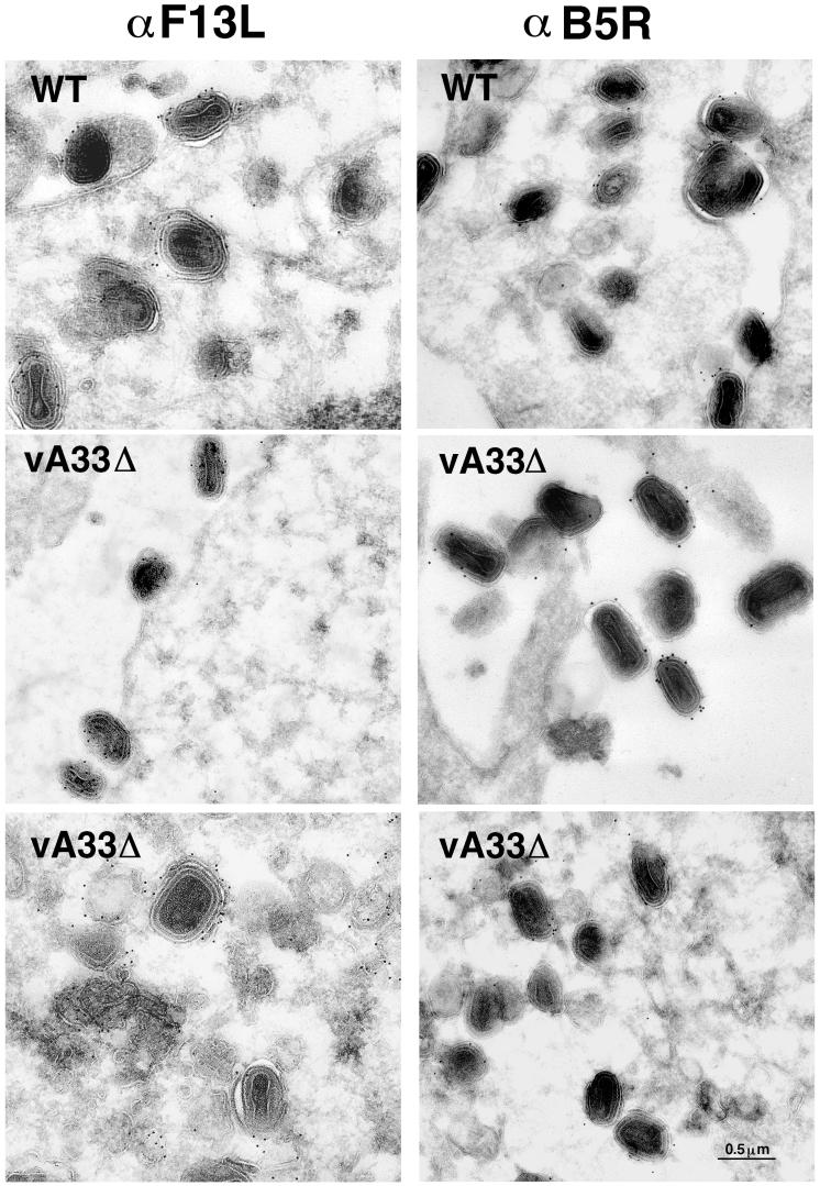
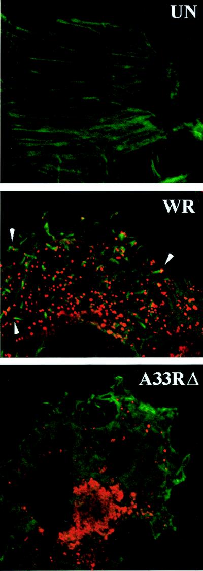
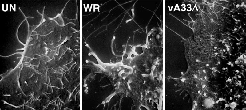
The vaccinia virus (VV) A33R gene encodes a highly conserved 23- to 28-kDa glycoprotein that is specifically incorporated into the viral outer envelope. The protein is expressed early and late after infection, consistent with putative early and late promoter sequences. To determine the role of the protein, two inducible A33R mutants were constructed, one with the late promoter and one with the early and late A33R promoter elements. Decreased A33R expression was associated with small plaques that formed comets in liquid medium. Using both an antibiotic resistance gene and a color marker, an A33R deletion mutant, vA33delta, was isolated, indicating that the A33R gene is not essential for VV replication. The plaques formed by vA33delta, however, were tiny, indicating that the A33R protein is necessary for efficient cell-to-cell spread. Rescue of the large-plaque phenotype was achieved by inserting a new copy of the A33R gene into the thymidine kinase locus, confirming the specific genetic basis of the phenotype. Although there was a reduction in intracellular virus formed in cells infected with vA33delta, the amount of infectious virus in the medium was increased. The virus particles in the medium had the buoyant density of extracellular enveloped viruses (EEV). Additionally, amounts of vA33delta cell-associated extracellular enveloped viruses (CEV) were found to be normal. Immunogold electron microscopy of cells infected with vA33delta demonstrated the presence of the expected F13L and B5R proteins in wrapping membranes and EEV; however, fully wrapped vA33delta intracellular enveloped viruses (IEV) were rare compared to partially wrapped particles. Specialized actin tails that propel IEV particles to the periphery and virus-tipped microvilli (both common in wild-type-infected cells) were absent in cells infected with vA33delta. This is the first deletion mutant in a VV envelope gene that produces at least normal amounts of fully infectious EEV and CEV and yet has a small-plaque phenotype. These data support a new model for VV spread, emphasizing the importance of virus-tipped actin tails.


Figures

FIG. 1
FIG. 1
Representation of the genomes of A33R inducible and deletion mutants. In the top row, relevant features of the genome of WT-A33 (vlacI) are shown. The A32L, A33R, and A34R ORFs are represented by ovals, and the tandem early and late promoter elements of the A33R gene are shown boxed. Arrows indicate the directions of transcription. In the second row, the genome of vA33full is represented. The locations of the E. coli gpt gene and lacO are indicated. The vA33late mutant, depicted in the third row, is similar to vA33full except that the A33R early promoter has been deleted. The last row represents part of the genome of vA33Δ with the neo/GUS genes replacing A33R.
FIG. 2
FIG. 2
Appearance of plaques formed by A33R inducible mutants. BS-C-1 cell monolayers were infected with vA33late, vA33full, or WT-A33 virus in the absence (top row) or presence (bottom row) of IPTG. After 29 h, the medium was removed and the monolayers were fixed and stained with 0.1% crystal violet in 20% ethanol.
FIG. 3
FIG. 3
Synthesis of A33R protein. BS-C-1 cells were infected with vA33late, vA33full, or WT-A33 virus with IPTG or AraC as indicated. After 2, 4, 6, or 12 h, the medium was removed, the cells were incubated for 1 h with [35S]methionine, and the cells were lysed. The A33R protein was immunoprecipitated with MAb 4, resolved by sodium dodecyl sulfate-polyacrylamide gel electrophoresis, and autoradiographed. The molecular weights (MW; 103) of markers are shown on the left. The arrows on the right point to the A33R protein.
FIG. 4
FIG. 4
Appearance of plaques formed by the A33R deletion mutant. BS-C-1 or RK13 monolayers were infected with WT-A33, vA33Δ, or vResA33. After 48 h, the medium was removed and the monolayers were fixed and stained with 0.1% crystal violet in 20% ethanol. At least 30 infectious foci or plaques are present in each well shown.
FIG. 5
FIG. 5
Yields of extracellular and cell-associated infectious virus. RK13 cells were infected with WT-A33 or vA33Δ at a multiplicity of 10. The inocula were removed after 2 h and replaced with fresh medium. At the indicated times, the medium was removed and centrifuged to pellet detached cells. Adherent cells were scraped into 1 ml of fresh medium and combined with pelleted cells, frozen, and thawed three times, and sonicated for 30 s. The titers of the virus from the medium and the cells were determined in duplicate on BS-C-1 cell monolayers.
FIG. 6
FIG. 6
Purification of wrapped and unwrapped virus particles by buoyant density centrifugation. [35S]methionine-labeled WT-A33 or vA33Δ virus was harvested from the medium, the lysed cells (intracellular), or intact, washed cells that were trypsin treated to release virus attached to the cell surface. Virus samples were centrifuged in CsCl gradients, and the positions of wrapped (wrap) and unwrapped (un) particles are indicated with arrows.
FIG. 7
FIG. 7
Induction of syncytia by WT-A33 or vA33Δ. BS-C-1 cells were infected for 12 h, treated for 2 min with buffer at pH 5.5 or 7.4, and then returned to the growth medium for 5 h and examined by phase-contrast microscopy.
FIG. 8
FIG. 8
Electron micrographs of Epon-embedded infected cells. RK13 cells were infected with WT-A33 or vA33Δ. After 24 h, the cells were fixed in glutaraldehyde and embedded in Epon. In WT-A33-infected cells (left panel), the arrow indicates an example of a fully wrapped IEV particle. In vA33Δ-infected cells (middle and right panels) arrows indicate partially wrapped IMV (incomplete IEV). It is important to note the numerous membrane vesicles with associated virus particles in the middle panel. Bars, 0.5 μm.
FIG. 9
FIG. 9
Immunogold labeling of viral membranes. RK13 cells were infected with WT-A33 or vA33Δ for 24 h, fixed in paraformaldehyde, cryosectioned, and incubated with antibodies to the F13L or B5R EEV-specific proteins and then protein A-gold.
FIG. 10
FIG. 10
Detection of actin filaments in VV-infected cells. HeLa cells were left uninfected (UN) or were infected with WT VV (strain WR) or vA33Δ. At 16 h, the cells were fixed, permeabilized, and incubated with FITC-conjugated phalloidin and a rabbit polyclonal serum that recognizes the B5R and F13L proteins, followed by rhodamine-conjugated antirabbit antibody. The images were examined by confocal microscopy.
FIG. 11
FIG. 11
Scanning electron microscopy of cells infected with VV. HeLa cells were left uninfected (UN) or were infected at a multiplicity of 10 with WT VV (strain WR) or vA33Δ. After 17 h, the cells were fixed with glutaraldehyde and the samples were coated with gold-palladium alloy and viewed with an Amray 1820D microscope.

References

    1. Appleyard G, Hapel A J, Boulter E A. An antigenic difference between intracellular and extracellular rabbitpox virus. J Gen Virol. 1971;13:9–17. - PubMed
    1. Blasco R, Moss B. Extracellular vaccinia virus formation and cell-to-cell virus transmission are prevented by deletion of the gene encoding the 37,000-dalton outer envelope protein. J Virol. 1991;65:5910–5920. - PMC - PubMed
    1. Blasco R, Moss B. Role of cell-associated enveloped vaccinia virus in cell-to-cell spread. J Virol. 1992;66:4170–4179. - PMC - PubMed
    1. Blasco R, Sisler J R, Moss B. Dissociation of progeny vaccinia virus from the cell membrane is regulated by a viral envelope glycoprotein: effect of a point mutation in the lectin homology domain of the A34R gene. J Virol. 1993;67:3319–3325. - PMC - PubMed
    1. Boulter E A, Appleyard G. Differences between extracellular and intracellular forms of poxvirus and their implications. Prog Med Virol. 1973;16:86–108. - PubMed

MeSH terms

LinkOut - more resources