. 1998 May;72(5):4237-42.
doi: 10.1128/JVI.72.5.4237-4242.1998.

Detection of a novel bovine lymphotropic herpesvirus

Affiliations

Detection of a novel bovine lymphotropic herpesvirus

J Rovnak et al. J Virol. 1998 May.

Abstract

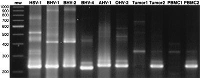
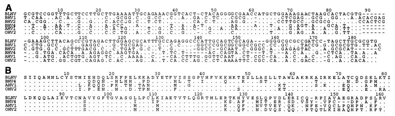
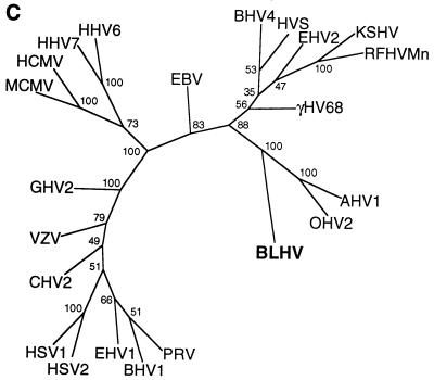
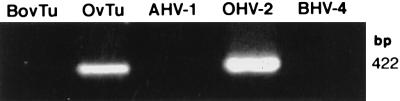
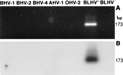
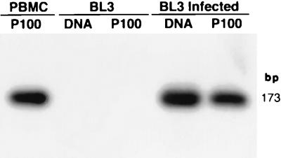
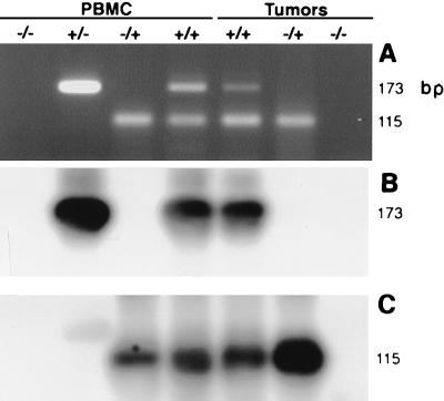
Degenerate PCR primers which amplify a conserved region of the DNA polymerase genes of the herpesvirus family were used to provide sequence evidence for a new bovine herpesvirus in bovine B-lymphoma cells and peripheral blood mononuclear cells (PBMC). The sequence of the resultant amplicon was found to be distinct from those of known herpesvirus isolates. Alignment of amino acid sequences demonstrated 70% identity with ovine herpesvirus 2, 69% with alcelaphine herpesvirus 1, 65% with bovine herpesvirus 4, and 42% with bovine herpesvirus 1. Phylogenetic analysis placed this putative virus within the tumorigenic Gammaherpesvirinae subfamily, and it is tentatively identified as bovine lymphotropic herpesvirus. This novel agent was expressed in vitro from infected PBMC, and cell-free supernatants were used to transfer infection to a bovine B-cell line, BL3. Analysis, with specific PCR primers, of DNA from bovine PBMC and lymphoma cells identified infection in blood of 91% of adult animals (n = 101), 63% of lymphomas (n = 32), and 38% of juveniles (n = 13). Of the adults, herpesvirus infection was present in 94% of animals that were seropositive for bovine leukemia virus (BLV) (n = 63) and in 87% of BLV-seronegative animals (n = 38). Of the seropositive group, 17 animals exhibited persistent lymphocytosis, and 100% of these were herpesvirus positive by PCR. A role for bovine lymphotropic herpesvirus as a cofactor in BLV pathogenesis is considered.


Figures

FIG. 1
FIG. 1
Gel analysis of products of degenerate herpesvirus primers. BHV1, BHV2, BHV4, and AHV1 were amplified from viral DNA preparations. HSV1-, OHV2-, and BLV-negative (Tumor1) and -positive (Tumor2) bovine tumors and BLV-negative (PBMC1) and -positive (PBMC2) PBMC were amplified from 1 μg of cellular DNA. Position of molecular weight markers (mw) are indicated in base pairs.
FIG. 2
FIG. 2
Analysis of DNA polymerase sequences of bovine herpesviruses. (A) Alignment of DNA sequences excluding TGV- and IYG-primed regions. The sequence obtained from bovine tumor is designated BLHV. Sequences for AHV1, BHV2, BHV4, OHV2, and BLHV were derived from the amplicons shown in Fig. 1. The sequence for BHV1 was obtained from GenBank (accession no. emb Z78205) (B) Alignment of amino acid sequences of gammaherpesviruses of cattle. Sequence corresponds to 478-bp of DNA internal to the DFA- and IYG-primed regions. Additional BLHV, BHV4, and OHV2 upstream sequences were obtained from PCR amplicons. Upstream AHV1 sequence was obtained from GenBank (accession no. AF005370) (C) Phylogenetic tree resulting from analysis of sequences in panel B and additional herpesvirus sequences from GenBank: CHV1 (canine herpesvirus 1; accession no. emb X89500), EBV (HHV4; accession no. V01555), EHV1 (equine herpesvirus 1; accession no. M86664), EHV2 (accession no. U20824), γHV68 (murine gammaherpesvirus 68; accession no. U97553), HCMV (human cytomegalovirus, HHV5; accession no. M14709), HHV6 (accession no. emb X83413), HHV7 (accession no. U43400), HSV1 (accession no. emb X04771), HSV2 (HHV2; accession no. M16321), HVS (saimiriine herpesvirus 2; accession no. M31122), KSHV (accession no. U93872), GHV2 (Marek’s disease virus, gallid herpesvirus 2; accession no. L40431), MCMV (murine cytomegalovirus; accession no. U68299), PRV (pseudorabies virus; accession no. L24487), RFHVMn (retroperitoneal fibromatosis herpesvirus of Macaca nemestrina; accession no. AF005478), and VZV (varicella-zoster virus, HHV3; accession no. emb X04370).
FIG. 2
FIG. 2
Analysis of DNA polymerase sequences of bovine herpesviruses. (A) Alignment of DNA sequences excluding TGV- and IYG-primed regions. The sequence obtained from bovine tumor is designated BLHV. Sequences for AHV1, BHV2, BHV4, OHV2, and BLHV were derived from the amplicons shown in Fig. 1. The sequence for BHV1 was obtained from GenBank (accession no. emb Z78205) (B) Alignment of amino acid sequences of gammaherpesviruses of cattle. Sequence corresponds to 478-bp of DNA internal to the DFA- and IYG-primed regions. Additional BLHV, BHV4, and OHV2 upstream sequences were obtained from PCR amplicons. Upstream AHV1 sequence was obtained from GenBank (accession no. AF005370) (C) Phylogenetic tree resulting from analysis of sequences in panel B and additional herpesvirus sequences from GenBank: CHV1 (canine herpesvirus 1; accession no. emb X89500), EBV (HHV4; accession no. V01555), EHV1 (equine herpesvirus 1; accession no. M86664), EHV2 (accession no. U20824), γHV68 (murine gammaherpesvirus 68; accession no. U97553), HCMV (human cytomegalovirus, HHV5; accession no. M14709), HHV6 (accession no. emb X83413), HHV7 (accession no. U43400), HSV1 (accession no. emb X04771), HSV2 (HHV2; accession no. M16321), HVS (saimiriine herpesvirus 2; accession no. M31122), KSHV (accession no. U93872), GHV2 (Marek’s disease virus, gallid herpesvirus 2; accession no. L40431), MCMV (murine cytomegalovirus; accession no. U68299), PRV (pseudorabies virus; accession no. L24487), RFHVMn (retroperitoneal fibromatosis herpesvirus of Macaca nemestrina; accession no. AF005478), and VZV (varicella-zoster virus, HHV3; accession no. emb X04370).
FIG. 3
FIG. 3
Amplification of cellular and viral DNAs with OHV2-specific primers. BHV4 and AHV1 viral DNA preparations and 1 μg of DNA from BLHV-positive bovine tumor (BovTu), OHV2-positive ovine tumor (OvTu), and OHV2-positive control (OHV-2) were subjected to PCR with OHV2 tegument protein gene-specific primers 556 and 755, which yield a predicted 422-bp amplicon.
FIG. 4
FIG. 4
Amplification of viral and cellular DNAs with BLHV-specific primers. (A) Viral DNA preparations of BHV1, BHV2, BHV4, and AHV1 and cellular DNA from OHV2-positive cells and BLHV-positive (BLHV+) and -negative (BLHV) tumors were subjected to PCR with primers 5′BLHV and 3′BLHV, which yield a predicted 173-bp amplicon. (B) Southern blot of the gel in panel A hybridized with a nested, BLHV-specific probe.
FIG. 5
FIG. 5
Southern blot analysis of transfer of BLHV infection. DNA purified from material pelleted at 100,000 × g (P100) as well as cellular DNA (DNA) was amplified with BLHV-specific primers 5′BLHV and 3′BLHV, blotted, and hybridized with a nested, BLHV-specific probe. The first lane shows the result of amplification of DNA from material pelleted from BLHV-positive PBMC culture medium. The remaining lanes show the products of amplification of either cellular DNAs or pellets from medium of uninfected or infected BL3 cells.
FIG. 6
FIG. 6
Dual amplification of bovine PBMC and tumor DNAs with BLHV- and BLV-specific primers. (A) Gel analysis of samples which represent the variety of results for all 147 samples tested (BLHV and BLV). Samples were amplified from 1 μg of DNA or 15 μl of cell lysate (9 × 10⁴ mononuclear cells) with primer pairs 5′BLHV-3′BLHV and 5′LTR-3′LTR, which yield 173-bp BLHV and 115-bp BLV amplicons, respectively. (B) Southern blot of the gel in panel A hybridized with a nested, BLHV-specific probe. (C) Southern blot as in panel B stripped and reprobed with a BLV-specific probe.

References

    1. Albrecht J-C, Nicholas J, Biller D, Cameron K R, Biesinger B, Newman C, Wittman S, Craxton M A, Coleman H, Fleckenstein B, Honess R W. Primary structure of the herpesvirus saimiri genome. J Virol. 1992;66:5047–5058. - PMC - PubMed
    1. Bacon L D, Witter R L, Fadly A M. Augmentation of retrovirus-induced lymphoid leukosis by Marek’s disease herpesvirus in white leghorn chickens. J Virol. 1989;63:504–512. - PMC - PubMed
    1. Baxter S I F, Pow I, Bridgen A, Reid H W. PCR detection of the sheep-associated agent of malignant catarrhal fever. Arch Virol. 1993;132:145–159. - PubMed
    1. Cockerell G L, Rovnak J. The correlation between the direct and indirect detection of bovine leukemia virus infection in cattle. Leukocyte Res. 1988;12:465–469. - PubMed
    1. Degen J L, Neubauer M G, Degen S J, Seyfried C E, Morris D R. Regulation of protein synthesis in mitogen-activated bovine lymphocytes. Analysis of actin-specific and total mRNA accumulation and utilization. J Biol Chem. 1983;258:12153–12162. - PubMed

Publication types

MeSH terms

LinkOut - more resources