. 1998 May;72(5):4413-20.
doi: 10.1128/JVI.72.5.4413-4420.1998.

Bovine leukemia virus-induced lymphocytosis and increased cell survival mainly involve the CD11b+ B-lymphocyte subset in sheep

Affiliations

Bovine leukemia virus-induced lymphocytosis and increased cell survival mainly involve the CD11b+ B-lymphocyte subset in sheep

N Chevallier et al. J Virol. 1998 May.

Abstract

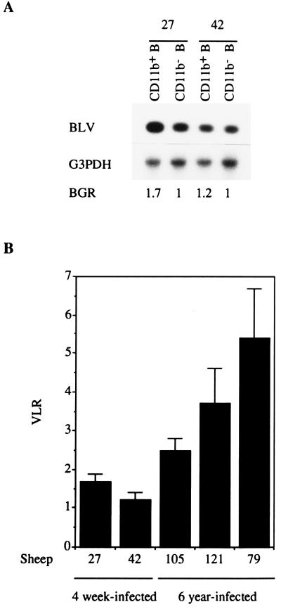
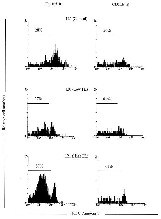
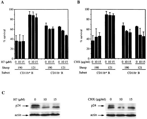
In this study, we show that bovine leukemia virus (BLV)-induced persistent lymphocytosis (PL) results from the in vivo expansion of the CD11b+ B-lymphocyte population. This subset shares phenotypic characteristics with murine and human B-1 cells. BLV interactions with the sheep B-1-like subset were explored. We found that B-1- and B-2-like cells are initially infected to similar extents. However, in long-term-infected sheep, the viral load is higher in B-1-like cells and only B-1- and not B-2-like cells show increased ex vivo survival compared to that in uninfected sheep. Ex vivo viral expression was found in both B-1- and B-2-like cells, indicating that both cell types support viral replication. Finally, cycloheximide and a protein kinase C inhibitor (H7) that blocks the ex vivo activation of viral expression did not affect the increased survival in B-1-like cells, suggesting that resistance to apoptosis is acquired in vivo. Collectively, these results indicate a peculiar susceptibility of sheep B-1-like cells to BLV transforming effects and further support the involvement of increased survival in BLV pathogenesis.


Figures

FIG. 1
FIG. 1
Expansion of the CD5+ and CD11b+ B-cell subsets in BLV-infected sheep. (A) The CD5 and CD21 markers were detected on PBMCs from a control sheep and a BLV-infected sheep with PL by incubating the cells with MAbs CC17 and DU2-104 followed by FITC-conjugated F(ab′)2 goat anti-mouse IgG1 (Caltag Laboratories, San Francisco, Calif.) and a phycoerythrin-conjugated F(ab′)2 goat anti-mouse IgM antibody (Jackson ImmunoResearch Laboratories, West Grove, Pa.). The percentages of cells in the different subsets are indicated. (B) Double labeling for CD11b (MAb CC125) and CD21 (MAb DU2-104) detection on PBMCs from the same sheep as for panel A. The percentages of cells in the different subsets are indicated.
FIG. 2
FIG. 2
BLV viral loads in CD11b+ and CD11b B cells in newly infected and long-term-infected sheep. (A) BLV provirus detection in sorted (by fluorescence-activated cell sorting) CD11b+ B and CD11b B cells from two 4-week-infected sheep (sheep 27 and 42). Uncultured CD11b+ and CD11b B cells were sorted, lysed, and analyzed for the detection of BLV provirus and the endogenous G3PDH gene by PCR, followed by Southern blotting and 32P-specific probing. The signals were analyzed with a PhosphorImager. The BGR obtained for each sorted population is shown. (B) VLRs for the BGRs of the CD11b+ and CD11b B-cell subsets were calculated for newly infected (4-week-infected) sheep and long-term (6-year)-infected sheep. Means and SEMs of the VLRs obtained for 8 (sheep 27 and 42) and 12 (sheep 105, 79, and 121) independent PCR experiments are shown.
FIG. 3
FIG. 3
CD11b+ B cells from BLV-infected sheep show increased ex vivo survival. PBMCs from control and BLV-infected sheep with low- and high-level PL were cultured for 48 h and labeled with FITC-annexin V and for detection of the CD11b and the CD21 markers [CC125 followed by a tricolor conjugated F(ab′)2 goat anti-mouse IgG1 antibody (Caltag Laboratories) and DU2-104 followed by a phycoerythrin-conjugated F(ab′)2 goat anti-mouse IgM antibody, respectively). The cells positive for both CD11b and CD21 (CD11b+) were gated, as were the cells positive for CD21 and negative for CD11b (CD11b). The proportions of surviving cells (annexin V negative) among the gated B cells are indicated.
FIG. 4
FIG. 4
In situ detection of BLV p24 expression in CD11b+ and CD11b B cells. Forty-eight-hour-cultured PBMCs from sheep 79 were labeled with an anti-CD21 MAb (DU2-104 followed by phycoerythrin-conjugated anti-mouse IgM) and an anti-CD11b MAb (ILA-130 followed by FITC-conjugated anti-mouse IgG2a), permeabilized with 70% methanol, and processed for BLV p24 detection (anti-p24 MAbs followed by triply conjugated anti-mouse IgG1). The p24-positive cells among gated CD11b+ and CD11b B cells are shown. The overlaid left histogram represents background fluorescence of permeabilized CD11b+ and CD11b B cells obtained with a control mouse IgG1 antibody to mutant p53 (Ctrl).
FIG. 5
FIG. 5
Inhibition of viral activation with H7 or CHX does not alter the increased survival in CD11b+ B cells from BLV-infected sheep with PL. PBMCs from a control sheep (sheep 190) and from a BLV-infected sheep with high-level PL (sheep 121) were cultured for 15 h without or with H7 (A) or with CHX (B). At the end of the culturing, the cells were labeled with FITC-annexin V and for detection of the CD11b (CC125) and CD21 (DU2-104) markers. The percentages of cell survival in each cell subset were obtained for three independent cultures, and means and standard deviations are reported. (C) Inhibition of viral capsid p24 expression with H7 and CHX treatments was analyzed by Western blotting with a pool of anti-BLV p24 MAbs (5 μg/ml) and an antiactin MAb (0.5 μg/ml, clone AC-15; Sigma) as an internal control.

References

    1. Birkebak T A, Palmer G H, Davis W C, Knowles D P, McElwain T F. Association of GP51 expression and persistent CD5+ B-lymphocyte expansion with lymphomatogenesis in bovine leukemia virus infected sheep. Leukemia. 1994;8:1890–1899. - PubMed
    1. Brauweiler A, Garrus J E, Reed J C, Nyborg J K. Repression of bax gene expression by the HTLV-1 Tax protein: implications for suppression of apoptosis in virally infected cells. Virology. 1997;231:135–140. - PubMed
    1. Burny A, Bruck C, Chanhenne H, Cleuter Y, Dekegel D, Ghysdael J, Kettmann R, Leclercq M, Leunen J, Mammerickx M, Portetelle D. Bovine leukemia virus: molecular biology and epidemiology. New York, N.Y: Raven Press; 1980. pp. 231–278.
    1. Cong Y-Z, Rabin E, Wortis H H. Treatment of murine CD5− B-cells with anti-Ig, but not LPS, induces surface CD5: two B-cell activation pathways. Int Immunol. 1991;3:467–476. - PubMed
    1. Cowan E P, Alexander R K, Daniel S, Kashanchi F, Brady J N. Induction of tumor necrosis factor alpha in human neuronal cells by extracellular human T-cell lymphotropic virus type 1 Tax1. J Virol. 1997;71:6982–6989. - PMC - PubMed

Publication types

Substances