. 1998 May;72(5):4434-41.
doi: 10.1128/JVI.72.5.4434-4441.1998.

Different patterns of neuronal infection after intracerebral injection of two strains of pseudorabies virus

Affiliations

Different patterns of neuronal infection after intracerebral injection of two strains of pseudorabies virus

J P Card et al. J Virol. 1998 May.

Abstract

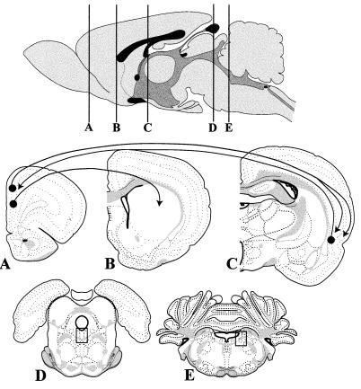
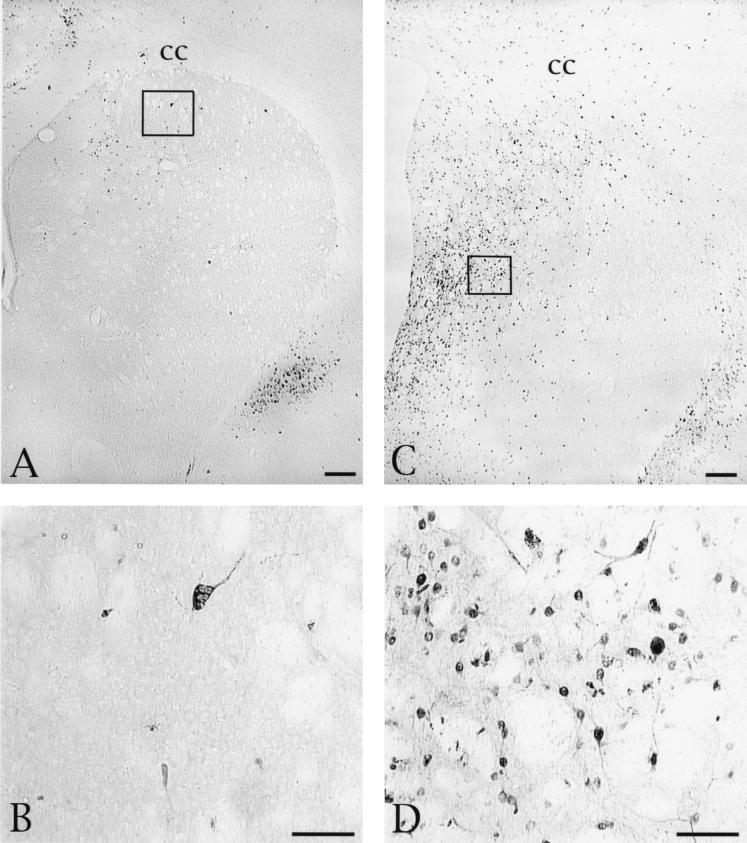
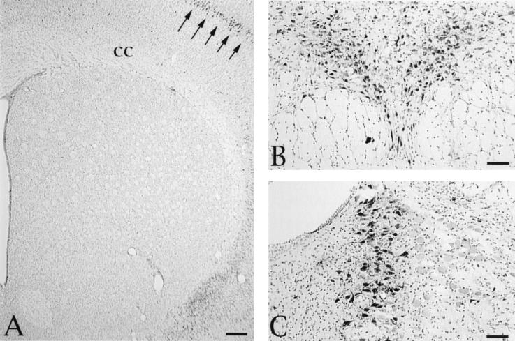
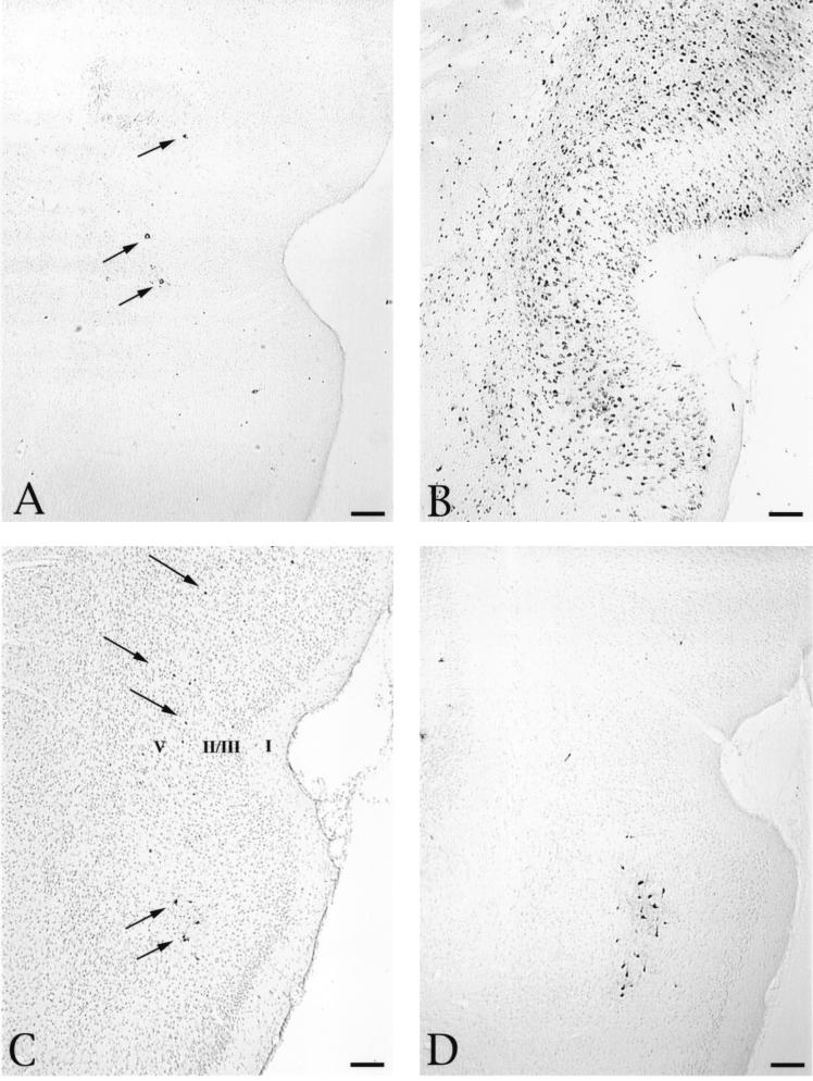
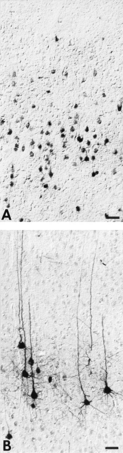
Pseudorabies virus (PRV), a swine neurotropic alphaherpesvirus, is known to invade the central nervous system (CNS) of a variety of animal species through peripherally projecting axons, replicate in the parent neurons, and then pass transsynaptically to infect other neurons of a circuit. Studies of the human pathogen herpes simplex virus type 1 have reported differences in the direction of transport of two strains of this virus after direct injection into the primate motor cortex. In the present study we examined the direction of transport of virulent and attenuated strains of PRV, utilizing injections into the rat prefrontal cortex to evaluate specific movement of virus through CNS circuitry. The data demonstrate strain-dependent patterns of infection consistent with bidirectional (anterograde and retrograde) transport of virulent virus and unidirectional (retrograde) transport of attenuated PRV from the site of injection. The distribution of infected neurons and the extent of transsynaptic passage also suggest that a release defect in the attenuated strain reduces the apparent rate of viral transport through neuronal circuitry. Finally, injection of different concentrations of virus influenced the onset of replication within a neural circuit. Taken together, these data suggest that viral envelope glycoproteins and virus concentration at the site of injection are important determinants of the rate and direction of viral transport through a multisynaptic circuit in the CNS.


Figures

FIG. 1
FIG. 1
Organization of the brain circuitry that was the subject of this analysis. Virus was injected into the PFC at level A. In the majority of cases the cannula tract passed vertically through intermediate layers of the anterior cingulate and prelimbic cortex; in a few instances the cannula extended further ventrally into the infralimbic cortex. All of these regions contain neurons that give rise to a dense axonal projection to the striatum (level B). This monosynaptic projection is not reciprocated, creating an ideal means of determining if the virus is being transported in an anterograde direction to produce a transsynaptic infection. The perirhinal cortex is among a large group of structures that maintain reciprocal connections with the PFC (level C). Neurons in the PFC give rise to axons that terminate in both superficial and deep layers of the perirhinal cortex. Therefore, an anterograde transsynaptic infection would be characterized by a multilaminar distribution of PRV-immunoreactive neurons. In contrast, the reciprocal projection of the perirhinal cortex to the PFC arises predominantly from lamina V. Therefore, retrograde transport of virus from the PFC would produce a selective infection of this layer of the perirhinal cortex. The boxed areas in levels D and E illustrate the location of the raphe nucleus and locus coeruleus illustrated in Fig. 3. See the text for further details on the organization of this circuitry. The templates used in this figure were modified from reference .
FIG. 2
FIG. 2
Distribution and morphology of striatal neurons infected by injection of PRV-Becker into the striatum. (A) Distribution of infected neurons in an animal sacrificed 27 h after injection of virus into the PFC. Scattered neurons are distributed across the dorsal and medial extent of the striatum in a pattern consistent with the established termination of afferents arising in the anterior cingulate and prelimbic cortex. (B) The boxed area of panel A is shown at higher magnification, demonstrating that large interneurons and smaller projection neurons are both infected at this survival interval. (C) Photomicrograph of another animal that was sacrificed 47 h after injection of virus into the PFC. A larger number of infected neurons are present in this animal, but the distribution is similar to that seen in the animal which survived for a shorter time and is consistent with the known distribution of PFC projections to the striatum. (D) The boxed area in panel C is shown at higher magnification, revealing viral immunoreactivity in the soma and processes of striatal neurons. Bars, 100 μm. cc, corpus callosum.
FIG. 3
FIG. 3
(A) Absence of anterograde transsynaptic infection of striatal neurons in an animal sacrificed 68 h following injection of 200 nl of PRV-Bartha into the PFC. Although infected neurons are clearly apparent in the overlying cortex (arrows), no PRV-immunoreactive cells are apparent in any region of the striatum. Other spatially distant regions with monosynaptic connections with the PFC also exhibited large numbers of infected neurons. Two of these areas in the brain stem are illustrated: dense viral immunoreactivity in the serotoninergic neurons of the dorsal raphe nucleus (B) and infected noradrenergic neurons in the locus coeruleus (C). These tissue sections were counterstained with cresyl violet to aid in the illustration of the cytoarchitecture in the regions of interest. Bars, 100 μm. cc, corpus callosum.
FIG. 4
FIG. 4
Distribution of infected neurons in the perirhinal cortex following injection of PRV-Becker or PRV-Bartha into the PFC. The distribution of infected neurons approximately 27 (A) and 48 (B) h after injection of 200 nl of PRV-Becker into the PFC is shown. At the shorter survival time, infected neurons are few and confined to lamina V of the perirhinal cortex (arrows). A substantially larger group of infected neurons are present in the perirhinal cortex 47 h after injection and extend through all cortical laminae. Scattered infection of layer V neurons is also observed 48 h after injection of 100 nl of either PRV-Becker (C) or PRV-Bartha (D) (arrows). The section illustrated in panel C was counterstained with cresyl violet to aid in the identification of cortical laminae, which are numbered according to the criteria defined by Swanson (43) to serve as points of orientation for defining the laminar disposition of infected neurons in the sections that were not counterstained. Bars, 200 μm.
FIG. 5
FIG. 5
Intracellular distribution of viral immunoreactivity in the perirhinal cortex 45 h after injection of PRV-Becker into the PFC (A) and 36 h after identical intracerebral injection of an equivalent concentration of PRV-Bartha (B). Viral antigens were identified with a rabbit polyclonal antibody generated against acetone-inactivated virus. The same dilution and period of incubation of tissue in the primary antibody was used in both cases, and the tissues were processed simultaneously. Note that immunoperoxidase reaction product is confined to the soma and primary dendrites of PRV-Becker-infected pyramidal neurons (A) whereas cells infected with PRV-Bartha exhibit a more extensive intracellular distribution of viral antigen, even though the animal was sacrificed 9 h earlier than the PRV-Becker-infected rat. Bars, 40 μm.

References

    1. Babic N, Mettenleiter T C, Flamand A, Ugolini G. Role of essential glycoproteins gII and gp50 in transneuronal transfer of pseudorabies virus from the hypoglossal nerves of mice. J Virol. 1993;67:4421–4426. - PMC - PubMed
    1. Babic N, Klupp B, Brack A, Mettenleiter T C, Ugolini G, Flamand A. Deletion of glycoprotein gE reduces the propagation of pseudorabies virus in the nervous system of mice after intranasal inoculation. Virology. 1996;219:279–284. - PubMed
    1. Barnett E M, Cassell M D, Perlman S. Two neurotropic viruses, herpes simplex virus type 1 and mouse hepatitis virus, spread along different neural pathways from the main olfactory bulb. Neuroscience. 1993;57:1007–1025. - PMC - PubMed
    1. Barnett E M, Evans G D, Sun N, Perlman S, Cassell M D. Anterograde tracing of trigeminal afferent pathways from the murine tooth pulp to cortex using herpes simplex virus type 1. J Neurosci. 1995;15:2972–2984. - PMC - PubMed
    1. Bartha A. Experimental reduction of virulence of Aujeszky’s disease virus. Magy Allatorv Lapja. 1961;16:42–45.

Publication types

Substances

LinkOut - more resources