. 1998 Apr;42(4):889-94.
doi: 10.1128/AAC.42.4.889.

Use of a rapid throughput in vivo screen to investigate inhibitors of eukaryotic topoisomerase II enzymes

Affiliations

Use of a rapid throughput in vivo screen to investigate inhibitors of eukaryotic topoisomerase II enzymes

T R Hammonds et al. Antimicrob Agents Chemother. 1998 Apr.

Abstract

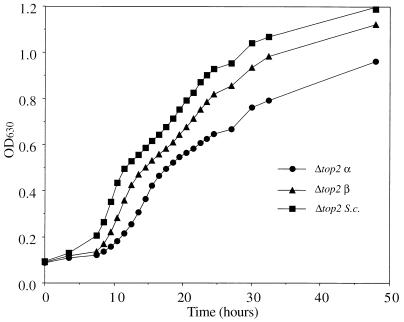
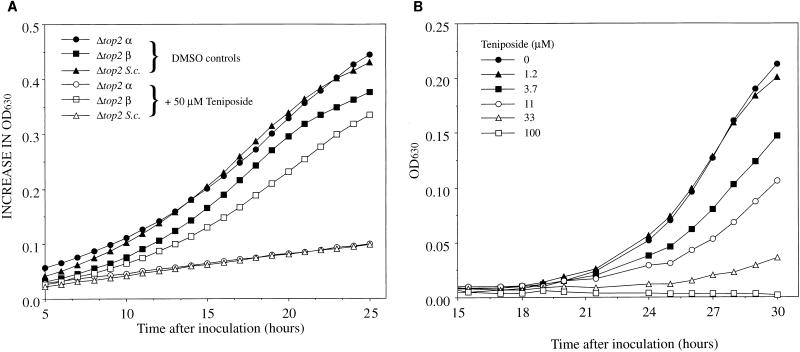
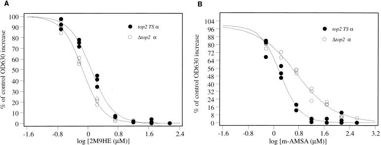
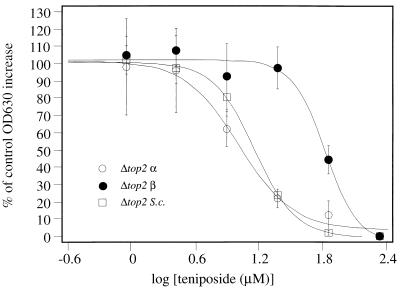
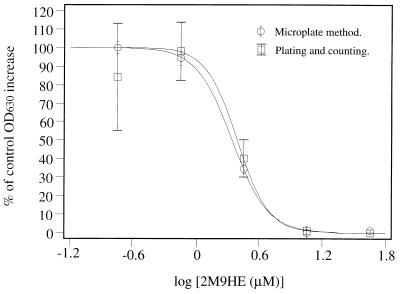
Topoisomerase II catalyzes the passage of one DNA helix through another via a transient double-stranded break. The essential nature of this enzyme in cell proliferation and its mechanism of action make it an ideal target for cytotoxic agents. Saccharomyces cerevisiae topoisomerase II has been frequently used as a model for testing potential inhibitors of eukaryotic topoisomerase II as antitumor agents. The standard in vivo method of estimating the sensitivity of S. cerevisiae to the antitopoisomerase drugs is via inhibition or kill curves which rely on viable-cell counts and is labor intensive. We present an alternative to this, a high-throughput in vivo screen. This method makes use of a drug-permeable S. cerevisiae strain lacking endogenous topoisomerase II, which is modified to express either human topoisomerase IIalpha or IIbeta or S. cerevisiae topoisomerase II carried on plasmids. Each modified strain expresses a full-length topoisomerase II enzyme, as opposed to the more commonly used temperature-sensitive S. cerevisiae mutant expressing yeast or yeast/human hybrid enzymes. A comparison of this new method with a plating-and-counting method gave similar drug sensitivity results, with increased accuracy and reduced manual input for the new method. The information generated has highlighted the sensitivities of different topoisomerase II enzymes and isoenzymes to several different classes of topoisomerase II inhibitor.


Figures

FIG. 1
FIG. 1
Growth of yeast cells in microwell plates as determined by monitoring OD630. Yeast cells were seeded into microplate wells and transferred to an incubator at 30°C as described in Materials and Methods. The OD630 was measured at 3- to 4-h intervals until significant increases were seen. The plate was then transferred to the reader at 30°C, and readings were taken hourly for 17 h. To conserve lamp time, readings were then taken at 2- to 5-h intervals.
FIG. 2
FIG. 2
Inhibition of yeast cell growth by teniposide. (A) Microplate wells were seeded as described in Materials and Methods with yeast strains only or with yeast strains plus 50 μM teniposide. After an initial lag phase the cells were transferred to a microplate reader at 30°C, and OD630 readings were taken at 30-min intervals. (B) Microplate wells were seeded as described in Materials and Methods with Δtop2 α. Teniposide was present in the media at the concentrations shown. The lag phase in these experiments is increased due to seeding at low levels. DMSO, dimethyl sulfoxide.
FIG. 3
FIG. 3
Dose-response curves of Δtop2 α and top2(Ts) α challenged with 2M9HE (A) and m-AMSA (B). Microplate wells were seeded as described in Materials and Methods, with one plate for each yeast strain. All experiments were carried out in triplicate. ID50s were obtained by calculation of the antilog of the x-axis value at the inflection point of each sigmoid-curve fit.
FIG. 4
FIG. 4
Dose-response curves of all Δtop2 yeast strains challenged with teniposide. Microplate wells were seeded as described in Materials and Methods, with one plate for each yeast strain. All experiments were carried out in triplicate. Values of the percentage of control growth are plotted as the averages of each set of three experiments, and error bars indicate one standard deviation.
FIG. 5
FIG. 5
Comparison of viable-count and microwell plate methods. Microplate wells were seeded as described in Materials and Methods, with Δtop2 α and 2M9HE in the media at five fourfold dilutions. All experiments, including control experiments, were performed in triplicate. The percentage increase in cell number relative to controls was estimated by viable-count and spectrophotometric methods. Error bars indicate one standard deviation.

Similar articles

Cited by

References

    1. Adachi N, Miyaike M, Kato S, Kanamaru R, Koyama H, Kikuchi A. Cellular distribution of mammalian DNA topoisomerase II is determined by its catalytically dispensable C-terminal domain. Nucleic Acids Res. 1997;25:3135–3142. - PMC - PubMed
    1. Berger J M, Gamblin S J, Harrison S C, Wang J C. Structure and mechanism of DNA topoisomerase-II. Nature. 1996;379:225–232. - PubMed
    1. Chen A Y, Liu L F. DNA topoisomerases: essential enzymes and lethal targets. Annu Rev Pharmacol Toxicol. 1994;34:191–218. - PubMed
    1. Cornarotti M, Tinelli S, Willmore E, Zunino F, Fisher L M, Austin C A, Capranico G. Drug sensitivity and sequence specificity of human recombinant DNA topoisomerases II alpha (p170) and II beta (p180) Mol Pharmacol. 1996;50:1463–1471. - PubMed
    1. Coutts J, Plumb J A, Brown R, Keith W N. Expression of topoisomerase-II-alpha and topoisomerase-II-beta in an adenocarcinoma cell-line carrying amplified topoisomerase-II-alpha and retinoic acid receptor-alpha genes. Br J Cancer. 1993;68:793–800. - PMC - PubMed

Publication types

MeSH terms

LinkOut - more resources