. 1998 Apr 28;95(9):4831-6.
doi: 10.1073/pnas.95.9.4831.

Structure of the catalytic domain of avian sarcoma virus integrase with a bound HIV-1 integrase-targeted inhibitor

Affiliations

Structure of the catalytic domain of avian sarcoma virus integrase with a bound HIV-1 integrase-targeted inhibitor

J Lubkowski et al. Proc Natl Acad Sci U S A. .

Abstract

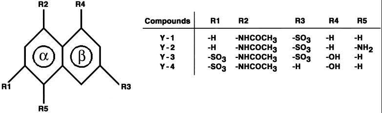
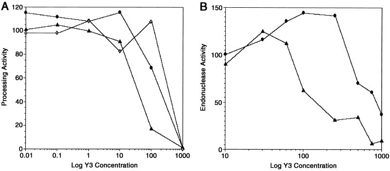
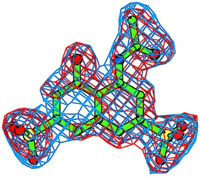
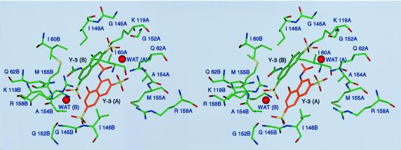
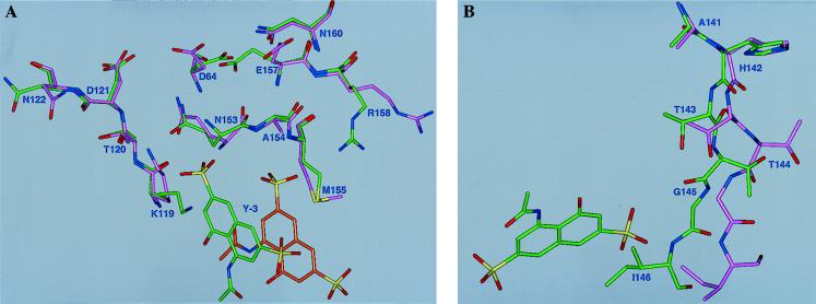
The x-ray structures of an inhibitor complex of the catalytic core domain of avian sarcoma virus integrase (ASV IN) were solved at 1.9- to 2.0-A resolution at two pH values, with and without Mn2+ cations. This inhibitor (Y-3), originally identified in a screen for inhibitors of the catalytic activity of HIV type 1 integrase (HIV-1 IN), was found in the present study to be active against ASV IN as well as HIV-1 IN. The Y-3 molecule is located in close proximity to the enzyme active site, interacts with the flexible loop, alters loop conformation, and affects the conformations of active site residues. As crystallized, a Y-3 molecule stacks against its symmetry-related mate. Preincubation of IN with metal cations does not prevent inhibition, and Y-3 binding does not prevent binding of divalent cations to IN. Three compounds chemically related to Y-3 also were investigated, but no binding was observed in the crystals. Our results identify the structural elements of the inhibitor that likely determine its binding properties.


Figures

Figure 1
Figure 1
Inhibitor compounds. A general formula is shown at left. The aromatic rings of the naphthalene system are marked as α and β. Definitions of groups R1–R5 are shown at right.
Figure 2
Figure 2
Inhibition of processing and endonuclease activities of ASV IN proteins by Y-3. (A) Processing activity of ASV IN (1–286) measured using Mn2+ (circles), Mg2+ (open diamonds), or Zn2+(triangles) as cofactor, with increasing concentrations of Y-3. Reactions containing Mn2+ were incubated at 37°C for 10 min.; those containing either Mg2+ or Zn2+ were incubated for 60 min. Processing activity is expressed as relative percentage of positive (no inhibitor) control. (B) Endonucleolytic activity of ASV IN (52–207) was measured using Mn2+(circles) or Zn2+(triangles), with increasing concentrations of Y-3. Reactions containing Mn2+ were incubated as above; those containing Zn2+ were incubated for 120 min. Data are expressed as in A.
Figure 3
Figure 3
An electron density (ED) map for the inhibitor Y-3. The initial Fo–Fc ED map is contoured at the 2.5σ level (red); the final 2Fo-Fc ED map is contoured at the 1.1σ level (blue). The refined conformation of the Y-3 model is shown in green. Prepared with bobscript, a modification of molscript (36).
Figure 4
Figure 4
Stereoview of the Y-3 binding site, shown approximately along the crystallographic twofold axis. Structural elements contributed by either monomer of the enzyme are labeled with A or B, respectively, after the residue number. Water molecules stabilizing the interactions between the two Y-3 molecules, WAT (A) and WAT (B), are shown.
Figure 5
Figure 5
Inhibitor binding site. Conformational differences between the Y-3- ASV IN (core) complex [pH 5.6 (green)] and the unliganded enzyme, Protein Data Bank code 1ASV (magenta). (A) Residues with altered conformation are labeled. For Arg 158, one of two alternate conformations is shown. The bound Y-3 molecule and its symmetry-related copy are both shown. (B) Conformational changes in the flexible loop sections (residues 141–146) with bound inhibitor molecules.

References

    1. Katz R A, Skalka A M. Annu Rev Biochem. 1994;63:133–173. - PubMed
    1. Vink C, Groeneger O A M, Plasterk R H. Nucleic Acids Res. 1993;21:1419–1425. - PMC - PubMed
    1. Goff S P. Annu Rev Genet. 1992;26:527–544. - PubMed
    1. Bujacz G, Jaskolski M, Alexandratos J, Wlodawer A, Merkel G, Katz R A, Skalka A M. Structure. 1996;4:89–96. - PubMed
    1. Bujacz G, Alexandratos J, Wlodawer A, Merkel G, Andrake M, Katz R A, Skalka A M. J Biol Chem. 1997;272:18161–18168. - PubMed

Publication types