. 1998 Apr 28;95(9):4876-81.
doi: 10.1073/pnas.95.9.4876.

Crystal structures of two tropinone reductases: different reaction stereospecificities in the same protein fold

Affiliations

Crystal structures of two tropinone reductases: different reaction stereospecificities in the same protein fold

K Nakajima et al. Proc Natl Acad Sci U S A. .

Abstract

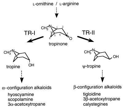
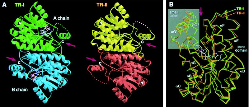
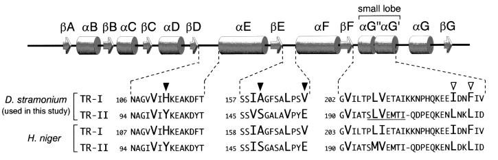
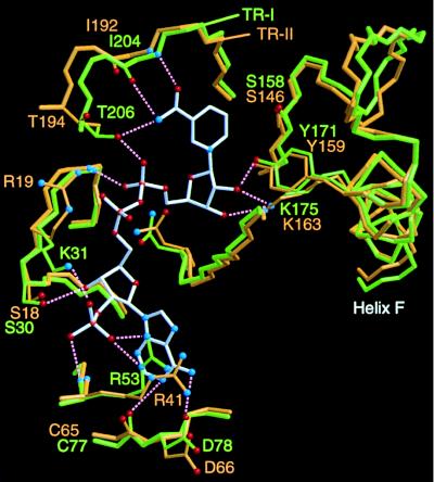
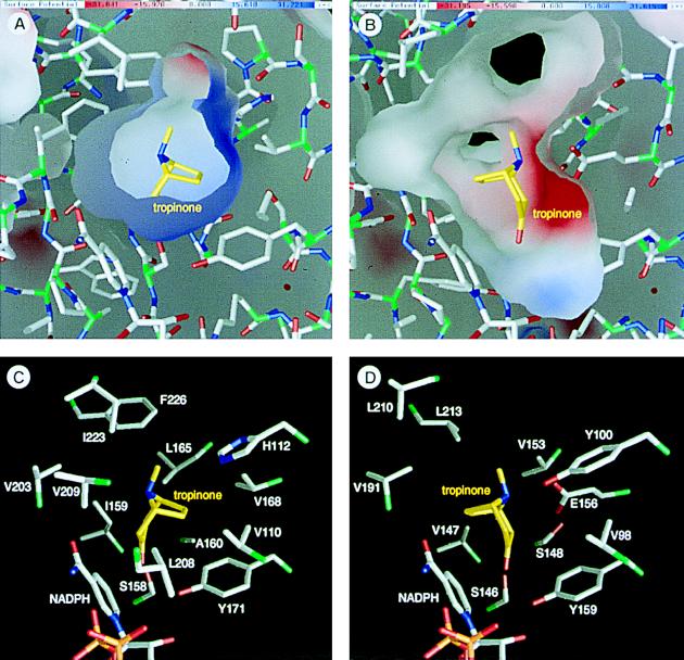
A pair of tropinone reductases (TRs) share 64% of the same amino acid residues and belong to the short-chain dehydrogenase/reductase family. In the synthesis of tropane alkaloids in several medicinal plants, the TRs reduce a carbonyl group of an alkaloid intermediate, tropinone, to hydroxy groups with different diastereomeric configurations. To clarify the structural basis for their different reaction stereospecificities, we determined the crystal structures of the two enzymes at 2.4- and 2.3-A resolutions. The overall folding of the two enzymes was almost identical. The conservation was not confined within the core domains that are conserved within the protein family but extended outside the core domain where each family member has its characteristic structure. The binding sites for the cofactor and the positions of the active site residues were well conserved between the two TRs. The substrate binding site was composed mostly of hydrophobic amino acids in both TRs, but the presence of different charged residues conferred different electrostatic environments on the two enzymes. A modeling study indicated that these charged residues play a major role in controlling the binding orientation of tropinone within the substrate binding site, thereby determining the stereospecificity of the reaction product. The results obtained herein raise the possibility that in certain cases different stereospecificities can be acquired in enzymes by changing a few amino acid residues within substrate binding sites.


Figures

Figure 1
Figure 1
Reactions catalyzed by TRs. TRs reduce the carbonyl group of a common substrate, tropinone, to hydroxy groups with different configurations. The reaction products tropine (3α-hydroxytropane) and ψ-tropine (3β-hydroxytropane) do not interconvert, rather they are further metabolized to various alkaloid products. Both enzymes require NADPH as the hydride donor. Carbon atoms of the tropane ring system are numbered in tropinone.
Figure 2
Figure 2
(A) Structures of TR dimers. Subunits of TR-I (green and blue) and TR-II (yellow and red) are related, respectively, by a noncrystallographic and crystallographic twofold axis positioned at the center of each dimer and oriented perpendicular to the plane of the figure. NADP+ bound in the TR-I subunits is shown by ball-and-stick models. Disordered regions in chain A of TR-I and both of the TR-II subunits are shown by dots. (B) Cα traces of TR-I (green) and TR-II (orange) subunits are superimposed by the program lsqkab (9) using all possible Cα pairs. The binding position of NADP+ in TR-I and the side chains of the three catalytic residues also are shown. The small lobes are shown in a gray background. Arrows indicate the clefts formed between the core domain and the small lobe (A and B). The figures were prepared by using the programs molscript (13) and raster3d (14).
Figure 3
Figure 3
Schematic representation of the secondary structural elements along the TR polypeptide. Amino acid sequence alignments are shown for the regions surrounding the putative tropinone binding residues. The sequences of the H. niger TRs are also shown for comparison. Amino acids predicted to be in contact with the tropinone substrate are shown in larger uppercase type, among which the positions with enzyme type-specific substitutions are indicated by arrowheads (solid arrowhead, substitution with polarity change; open arrowhead, substitution conserving hydrophobicity). The disordered TR-II polypeptide segment is underlined.
Figure 4
Figure 4
Superimposition of the region encompassing the cofactor binding site and catalytic residues of TRs. The binding position of NADP+ (bonds shown in white) was determined for TR-I. Bonds in the TR-I and TR-II proteins are shown in green and orange, respectively. For simplicity, only the protein side chains that directly interact with the cofactor atoms are shown. Possible hydrogen bonding interactions are indicated by dotted lines, although many other hydrogen bonding interactions through water molecules could be predicted from the structural model. Three catalytic residues (Ser, Tyr, Lys) are shown at the right. The figure was prepared by using the programs molscript (13) and raster3d (14).
Figure 5
Figure 5
Environments of the substrate binding sites and the binding models of tropinone. (A and B) Inner molecular surfaces of the tropinone binding pockets of TR-I (A) and TR-II (B). Electrostatic charge distributions are shown on the molecular surfaces in blue (positive charge) and red (negative charge). Note that the inner surface of TR-II (B) is somewhat deformed at the lower left corner due to the absence of several residues that constitute the small lobe. (C and D) Amino acid side chains that form the tropinone binding pockets viewed from the same direction as in A and B. Two TR-II residues (D) corresponding to the Leu-208 and Val-209 of TR-I (C) are missing in this structural model. Bonds from the carbon atoms in tropinone are shown in yellow, whereas the protein bonds from the Cα atoms are green to half their lengths. This figure was prepared by the grasp program (23).

References

    1. Hashimoto T, Nakajima K, Ongena G, Yamada Y. Plant Physiol. 1992;100:836–845. - PMC - PubMed
    1. Nakajima K, Hashimoto T, Yamada Y. Proc Natl Acad Sci USA. 1993;90:9591–9595. - PMC - PubMed
    1. Nakajima K, Hashimoto T, Yamada Y. Plant Physiol. 1993;103:1465–1466. - PMC - PubMed
    1. Jörnvall H, Persson B, Krook M, Atrian S, Gonzàlez-Duarte R, Jeffery J, Ghosh D. Biochemistry. 1995;34:6003–6013. - PubMed
    1. Nakajima K, Hashimoto T, Yamada Y. J Biol Chem. 1994;269:11695–11698. - PubMed

Publication types

LinkOut - more resources