. 1998 Apr 28;95(9):4888-93.
doi: 10.1073/pnas.95.9.4888.

Mechanism of platelet inhibition by nitric oxide: in vivo phosphorylation of thromboxane receptor by cyclic GMP-dependent protein kinase

Affiliations

Mechanism of platelet inhibition by nitric oxide: in vivo phosphorylation of thromboxane receptor by cyclic GMP-dependent protein kinase

G R Wang et al. Proc Natl Acad Sci U S A. .

Abstract

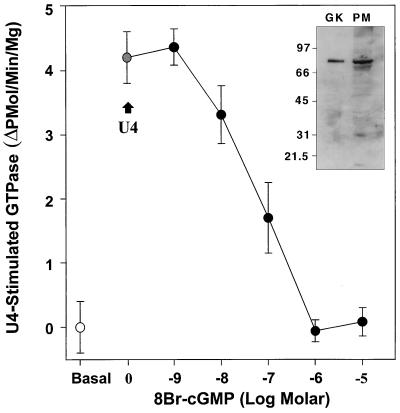
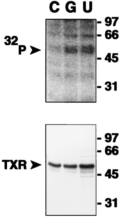
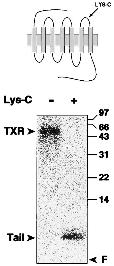
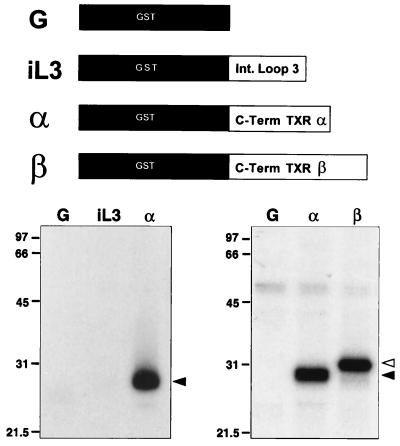
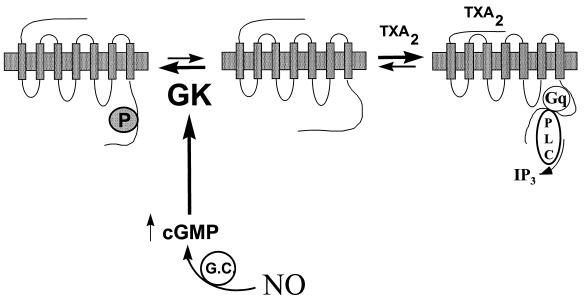
Nitric oxide (NO) is a potent vasodilator and inhibitor of platelet activation. NO stimulates production of cGMP and activates cGMP-dependent protein kinase (G kinase), which by an unknown mechanism leads to inhibition of Galphaq-phospholipase C-inositol 1, 4,5-triphosphate signaling and intracellular calcium mobilization for several important agonists, including thromboxane A2 (TXA2). To explore the mechanism of platelet inhibition by NO, activation of platelet TXA2 receptors in the presence of cGMP was studied. The nonhydrolyzable analog 8-bromo-cyclic GMP (8-Br-cGMP) potently inhibited activation of the TXA2-specific GTPase in platelet membranes in a concentration-dependent fashion, suggesting that G kinase catalyzes the phosphorylation of some proximal component of the receptor-G protein signaling pathway. Nanomolar concentrations of G kinase were found to catalyze the phosphorylation of platelet TXA2 receptors in vitro, but not Galphaq copurifying with the TXA2 receptors in these experiments. Using immunoaffinity methods, in vivo phosphorylation of TXA2 receptors by cyclic GMP was demonstrated from 32P-labeled cells treated with 8-Br-cGMP. Peptide mapping studies of in vivo phosphorylated TXA2 receptors demonstrated cGMP mediates phosphorylation of the carboxyl terminus of the TXA2 receptor. G kinase also catalyzed the phosphorylation of peptides corresponding to the cytoplasmic tails of both alpha and beta forms of the receptor but not control peptide or a peptide corresponding to the third intracytoplasmic loop of the TXA2 receptor. These data identify TXA2 receptors as cGMP-dependent protein kinase substrates and support a novel mechanism for the inhibition of cell function by NO in which activation of G kinase inhibits signaling by G protein-coupled receptors by catalyzing their phosphorylation.


Figures

Figure 1
Figure 1
TXA2 receptor-coupled GTPase activation in human platelet membranes is inhibited by 8-Br-cGMP. Platelet membranes were stimulated with 10 μM of the TXA2 analog U46619 in the absence or presence of varying concentrations of 8-Br-cGMP. Basal GTPase activity (open circle) and U46619-stimulated GTPase activity in buffer-treated membranes (grey circle; +U4) are shown, as is the progressive inhibition of U46619-stimulated GTPase by 8-Br-cGMP (dark circles). The IC50 for inhibition of the TXA2-stimulated GTPase by 8-Br-cGMP is 75 nM (n = three experiments in triplicate). (Inset) Immunoblot demonstrating the presence of high levels of G kinase in the platelet membranes used for these studies. Lane 1, 0.2 μg of purified G kinase; lane 2, 79 μg of total platelet membrane protein.
Figure 2
Figure 2
Immunoblot demonstrating immunoaffinity isolation of thromboxane receptors: (Left) Western blot demonstrating isolation of TXA2 receptors from human platelet lysates. TXA2 receptors were isolated by incubating platelet lysates with TXR2 anti-receptor antibody precoupled to agarose beads. Bead eluates (E1–E3) were resolved by SDS/PAGE, transferred to nitrocellulose, and then immunoblotted with TXR2 antibody. L, platelet lysate; L*, lysate after incubating with TXR2 antibody beads; B*, IgG from TXR2 beads; E1–E3, sequential eluates from the TXR2 antibody beads incubated with platelet lysate, washed, and then eluted with low pH glycine buffer. The 47–53-kDa band in lanes L, E1, E2, and E3 is the platelet TXA2 receptor. (Right) Separate experiments demonstrating the expected (ref. 42) small decrease in Mr of the TXA2 receptor following endoproteinase Lys-C digestion. One of four similar experiments. R, TXA2 receptors; LC, receptor digested with endoproteinase Lys-C.
Figure 3
Figure 3
In vivo phosphorylation of immunopurified TXA2 receptor by cyclic GMP and the thromboxane analog U46619. TXA2 receptors were isolated by immunoaffinity methods from 32P-labeled HEL cells and resolved on SDS/PAGE gels, transferred to nitrocellulose, and then subjected to autoradiography and immunoblotting. C, vehicle control, 10 min; G, 10 mM 8-Br-cGMP, 15 min; U, 5μM U46619, 10 min. The lower panel is an immunoblot of the nitrocellulose shown phosphorylated in the upper panel with anti-TXA2 receptor antibody. One of five similar experiments is shown.
Figure 4
Figure 4
Endoproteinase Lys-C digestion of in vivo phosphorylated TXA2 receptors. Undigested in vivo phosphorylated TXA2 receptors (−) or receptors enzymatically digested by endoproteinase Lys-C (+) and resolved on 15% SDS/PAGE gel. Note that receptor-associated 32P is released by Lys-C digestion and migrates at 6–8 kDa, associated with the carboxyl-terminal tail fragment (“Tail”). F, gel front. One of two similar experiments is shown.
Figure 5
Figure 5
In vitro phosphorylation of peptide based on the C terminus of TXA2 receptors by G kinase. Kinase reactions contained 8 nM purified G kinase, [γ-32P]ATP, and recombinant GST protein (G), the GST-TXA2 receptor third intracytoplasmic loop (iL3), or the GST-TXA2 receptor tail sequence fusion protein corresponding to the α or β isoforms of the TXA2 receptor (α and β, respectively). Proteins were quantitated by Bradford assay and on Coomassie-stained gels, and the same input concentrations of protein were used for the various peptides. Left and right panels represent two different experiments. Reactions were resolved on 10% SDS/PAGE gels to generate the autoradiographs shown. (Right) Dark arrowhead denotes the TXA2 α carboxyl-terminal fusion protein; open arrowhead denotes the TXA2 receptor β carboxyl-terminal fusion protein. One of three similar experiments is shown in each case.
Figure 6
Figure 6
Proposed model for the inhibition of G protein-coupled receptor activation by nitric oxide and cyclic-GMP. Nitric oxide activates guanylate cyclase, producing cGMP and activating G kinase (GK). G kinase in turn phosphorylates the TXA2 receptor, which prevents or disrupts coupling of the receptor to its cognate GTP-binding protein Gq and thus inhibits activation of the effector, phospholipase C (PLC), preventing [Ca2+]i mobilization and cellular activation. The net effect is to shift the equilibrium in resting cells between precoupled receptor and uncoupled receptor toward the uncoupled state. It is also possible that the agonist-activated receptor is a target for G kinase. In this model, NO “sets the gain” for cellular activation by G protein-coupled receptors like the TXA2 receptor, resulting in a net decrease in platelet or vascular smooth muscle activation by physiologic agonists.

References

    1. Bredt D S, Snyder S H. Annu Rev Biochem. 1994;63:175–195. - PubMed
    1. Schafer A I, Alexander R W, Handin R I. Blood. 1980;55:649–654. - PubMed
    1. Mellion B T, Ignarro L J, Ohlstein E H, Pontecorvo E G, Hyman A L, Kadowitz P J. Blood. 1981;57:946–955. - PubMed
    1. Azuma H, Ishikawa M, Sekizaki S. Br J Pharmacol. 1986;88:411–415. - PMC - PubMed
    1. Mollace V, Salvemini D, Anggard E, Vane J. Br J Pharmacol. 1991;104:633–638. - PMC - PubMed

Publication types

MeSH terms

LinkOut - more resources