. 1998 Apr 28;95(9):4906-11.
doi: 10.1073/pnas.95.9.4906.

Endogenous cellulases in animals: isolation of beta-1, 4-endoglucanase genes from two species of plant-parasitic cyst nematodes

Affiliations

Endogenous cellulases in animals: isolation of beta-1, 4-endoglucanase genes from two species of plant-parasitic cyst nematodes

G Smant et al. Proc Natl Acad Sci U S A. .

Abstract

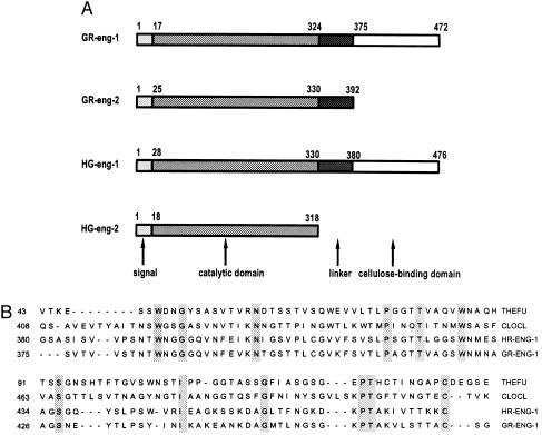
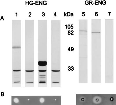
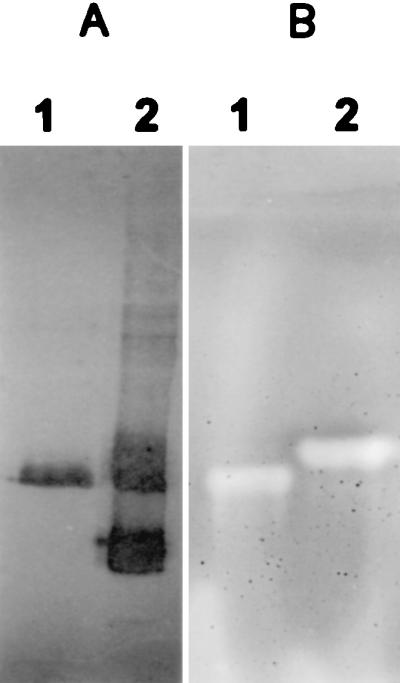
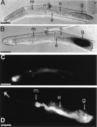
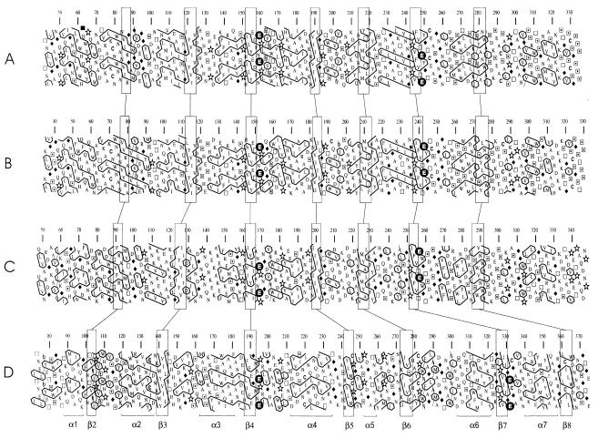
beta-1,4-Endoglucanases (EGases, EC 3.2.1.4) degrade polysaccharides possessing beta-1,4-glucan backbones such as cellulose and xyloglucan and have been found among extremely variegated taxonomic groups. Although many animal species depend on cellulose as their main energy source, most omnivores and herbivores are unable to produce EGases endogenously. So far, all previously identified EGase genes involved in the digestive system of animals originate from symbiotic microorganisms. Here we report on the synthesis of EGases in the esophageal glands of the cyst nematodes Globodera rostochiensis and Heterodera glycines. From each of the nematode species, two cDNAs were characterized and hydrophobic cluster analysis revealed that the four catalytic domains belong to family 5 of the glycosyl hydrolases (EC 3.2.1, 3.2.2, and 3.2.3). These domains show 37-44% overall amino acid identity with EGases from the bacteria Erwinia chrysanthemi, Clostridium acetobutylicum, and Bacillus subtilis. One EGase with a bacterial type of cellulose-binding domain was identified for each nematode species. The leucine-rich hydrophobic core of the signal peptide and the presence of a polyadenylated 3' end precluded the EGases from being of bacterial origin. Cyst nematodes are obligatory plant parasites and the identified EGases presumably facilitate the intracellular migration through plant roots by partial cell wall degradation.


Figures

Figure 1
Figure 1
(A) Global architecture of the four nematode β-1,4-endoglucanase precursor proteins. The amino acid positions of the functional domains are indicated above. (B) Sequence alignment of the C-terminal cellulose binding domains of G. rostochiensis ENG-1 and H. glycines ENG-1 with two representatives of the bacterial type of cellulose binding domains [Thermomonospora fusca EGase E-5 (THEFU), GenBank accession no. L01577, and Clostridium cellulovorans EGase D (CLOCL), GenBank accession no. M37434]. Residues conserved in all four cellulose binding domains are shaded.
Figure 2
Figure 2
Detection of recombinant nematode EGases in E. coli lysates on Western blot (A) and CMC hydrolysis assays (B). (A) Immunodetection was done with subventral gland specific mAbs (17). Lanes: 1, HG-ENG-1 produced from pET28c; 2, pET28c control; 3, HG-ENG-2 produced from pET28a; 4, pET28a control; 5, GR-ENG-1 from vector pMAL-c2; 6, GR-ENG-2 from vector pMAL-c2; 7, pMAL-c2 control. (B) Detection of CMCase activity (halo) in affinity-purified heterologous cyst nematode EGases that correspond to lanes above.
Figure 3
Figure 3
Comparative nonreducing SDS/PAGE of nematode homogenates following either Western blotting (A) or CMC overlay (B). (A) mAb MGR48 reacting with subventral gland protein of H. glycines (lane 1) and G. rostochiensis (lane 2). (B) Congo red stained agarose bed containing CMC showing hydrolysis activity (halo) by specific proteins of H. glycines (lane 1) and G. rostochiensis (lane 2).
Figure 4
Figure 4
Localization of EGase transcripts and proteins within cyst nematodes. (A and B) Anterior sections of preparasitic J2 of H. glycines hybridized with digoxigenin-labeled RNA probes transcribed from the HG-eng-2 cDNA. (A) No staining is observed with the sense probe. (B) Specific binding of the antisense probe to the posterior region of the subventral gland cells (g). The gland extensions (e) remain unstained. n, nucleolus of dorsal gland; m, metacorpal pump chamber. (C and D) Immunofluorescence of H. glycines using antiserum raised against recombinant nematode HG-ENG2. (C) No specific labeling with the preimmune serum. (D) Intense labeling of subventral glands with HG-ENG-2-specific immune serum. (Bar = 20 μm.)
Figure 5
Figure 5
Hydrophobic cluster analysis of the N-terminal catalytic domains of (A) HG-ENG-1, (B) GR-ENG-1, (C) EGase of B. subtilis (Swiss–Prot accession no. P07983), and (D) EGase A of C. cellulolyticum (Swiss–Prot accession no. P17901). The assignment of the secondary structure in C. cellulolyticum EGase A is according to Ducros et al. (29). The correspondence between the β strands in C. cellulolyticum EGase A and the equivalent strands in the other sequences is shown. The two catalytic residues in family 5 glycosyl hydrolases are shown as white letters in solid circles.

Comment in

References

    1. Carpita N C, Gibeaut D M. Plant J. 1993;3:1–30. - PubMed
    1. Beguin P, Aubert J. FEMS Microbiol Rev. 1994;13:25–58. - PubMed
    1. Teeri T T. Trends Biotechnol. 1997;15:160–167.
    1. Gilkes N R, Henrissat B, Kilburn D G, Miller R C, Jr, Warren R A. Microbiol Rev. 1991;55:303–315. - PMC - PubMed
    1. Klyosov A. Biochemistry. 1990;29:10577–10585. - PubMed

Publication types

Associated data

LinkOut - more resources