. 1998 Apr 28;95(9):4935-40.
doi: 10.1073/pnas.95.9.4935.

Specificity in cholesterol regulation of gene expression by coevolution of sterol regulatory DNA element and its binding protein

Affiliations

Specificity in cholesterol regulation of gene expression by coevolution of sterol regulatory DNA element and its binding protein

J N Athanikar et al. Proc Natl Acad Sci U S A. .

Abstract

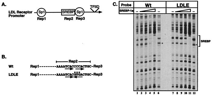
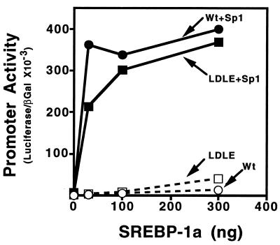
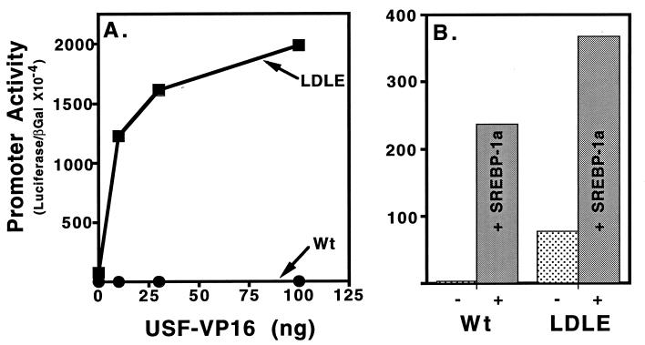
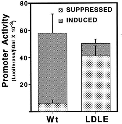
When demand for cholesterol rises in mammalian cells, the sterol regulatory element (SRE) binding proteins (SREBPs) are released from their membrane anchor through proteolysis. Then, the N-terminal region enters the nucleus and activates genes of cholesterol uptake and biosynthesis. Basic helix-loop-helix (bHLH) proteins such as SREBPs bind to a palindromic DNA sequence called the E-box (5'-CANNTG-3'). However, SREBPs are special because they also bind direct repeat elements called SREs. Importantly, sterol regulation of all promoters studied thus far is mediated by SREBP binding only to SREs. To study the reason for this we converted the direct repeat SRE from the sterol-regulated low-density lipoprotein receptor promoter into an E-box. In this report we show that SREBPs are still able to bind and activate this promoter however, sterol regulation is lost. The results are consistent with the mutant promoter being a target for promiscuous activation by constitutively expressed E-box binding bHLH proteins that are not regulated by cholesterol. Kim and coworkers [Kim, J. B., Spotts, G. D., Halvorsen, Y.-D., Shih, H.-M., Ellenberger, T., Towle, H. C. & Spiegelman, B. M. (1995) Mol. Cell. Biol. 15, 2582-2588] demonstrated that the dual DNA binding specificity of SREBPs is caused by a specific tyrosine in the conserved basic region of the DNA binding domain that corresponds to an arginine in all other bHLH proteins that recognize only E-boxes. Taken together the data suggest an evolutionary mechanism where a DNA binding protein along with its recognition site have coevolved to ensure maximal specificity and sensitivity in a crucial nutritional regulatory response.


Figures

Figure 1
Figure 1
Wild-type and mutant LDL receptor promoters: Structure and DNase I footprint analysis by SREBP-1a. A schematic view of the region of the LDL receptor promoter required for sterol regulation containing repeats 1–3 (A) and a more detailed view of the sequence of the wild-type (LDL) and mutant repeat 2 (LDLE) are shown (B). The mutated bases are indicated by ∗ and the direct repeat and palindromic motifs are indicated by the arrows. (C). DNA probes end-labeled with 32P on the top strand were prepared from plasmids containing the wild-type (Wt, lanes 1–6) or mutant (LDLE, lanes 7–12) promoters. DNA probe (0.01 pmol) were used in each binding reaction in the absence or presence of increasing amounts (0.02, 0.06, 0.2, and 0.6 pmol) of recombinant SREBP-1a (amino acids 1–490). DNase I footprinting was performed as described in Materials and Methods. The asterisk on the autoradiogram (C) indicates the position of differences in the observed cleavage pattern of free DNA due to the 3-bp difference between the two probes. The minimal size of the DNase I footprint is indicated by the bracket at the right of the figure.
Figure 2
Figure 2
Activation of the wild-type and mutant LDL receptor promoter by SREBP-1a and Sp1. Drosophila SL2 cells were transfected with increasing amounts of pPACSREBP-1a DNA alone (□, ○) or in the presence (▪, •) of a constant amount (25 ng/60-mm dish) of pPACSp1 expression vectors as indicated. The wild-type (Wt; •, ○) or mutant (LDLE; ▪, □) LDL receptor promoter fused to the luciferase gene (2 μg per 60-mm dish) were used as the reporters. Transfection, harvesting, preparation of cell extracts, and enzyme assays were performed as described in Materials and Methods. A pPAC-β-galactosidase expression vector (1 μg per 60-mm dish) was included in all transfections and its expression was used for normalization of the luciferase activity. This graph represents data from one of three individual experiments that showed similar results.
Figure 3
Figure 3
Activation of the wild-type and mutant LDL receptor promoter by USF2-VP16. (A). HepG2 cells were transfected with increasing amounts of plasmid cytomegalovirus-USF2-VP16 expression vector (abscissa) and wild-type (Wt; •) or mutant (LDLE; ▪) LDL receptor promoter luciferase reporter plasmids (5 μg per 60-mm dish) along with 5 μg per 60-mm dish of cytomegalovirus-β-galactosidase plasmid as indicated. (B) Companion dishes were transfected with the same reporter plasmids alone (−) or along with the SREBP-1a expression vector (+) as indicated (30 ng per 60-mm dish). Transfections were performed and analyzed as described in Materials and Methods and in the legend to Fig. 2.
Figure 4
Figure 4
Evaluation of sterol regulation for the wild-type and mutant LDL receptor promoters. The wild-type (Wt) or mutant (LDLE) LDL receptor promoter fused to the luciferase gene were transfected into CV-1 cells (5 μg per 60-mm dish) and 18 hr after transfection dishes were fed either medium containing lipoprotein-depleted serum (Induced) or medium containing lipoprotein-depleted serum plus 10 μg/ml of cholesterol and 1 μg/ml of 25-hydroxycholesterol (Suppressed). The cells were cultured for an additional 24 hr before harvest. The samples were processed as described in Materials and Methods. Each value represents the average of two independent experiments each performed in duplicate.

References

    1. Goldstein J, Brown M. Nature (London) 1990;343:425–430. - PubMed
    1. Osborne T F. Crit Rev Eukaryotic Gene Expression. 1995;5:317–335. - PubMed
    1. Brown M S, Goldstein J L. Cell. 1997;89:331–340. - PubMed
    1. Smith J R, Osborne T F, Brown M S, Goldstein J L, Gil G. J Biol Chem. 1988;263:18480–18487. - PubMed
    1. Osborne T F. J Biol Chem. 1991;266:13947–13951. - PubMed

Publication types

MeSH terms

LinkOut - more resources