

doi: 10.1073/pnas.95.9.4991.
Localization of nascent RNA and CREB binding protein with the PML-containing nuclear body
Affiliations
- PMID: 9560216
- PMCID: PMC20201
- DOI: 10.1073/pnas.95.9.4991
Item in Clipboard
Localization of nascent RNA and CREB binding protein with the PML-containing nuclear body
Proc Natl Acad Sci U S A.
.
Abstract
The cellular role of the PML-containing nuclear bodies also known as ND10 or PODs remains elusive despite links to oncogenesis and viral replication. Although a potential role in transcription has been considered, direct evidence has been lacking. By developing a novel in vivo nucleic acid labeling approach, we demonstrate the existence of nascent RNA polymerase II transcripts within this nuclear body. In addition, PML and the transactivation cofactor, CREB binding protein (CBP), colocalize within the nucleus. Furthermore, we show that CBP in contrast to PML is distributed throughout the internal core of the structure. Collectively, these findings support a role for this nuclear body in transcriptional regulation.
Figures

Confocal microscopy of HEp-2 cells microinjected with FITC–UTP. Fluorescent labeling was localized in PML-containing nuclear bodies and in coiled bodies, both of which are nuclear bodies of unknown function. Although not all PML bodies correspond to FITC–UTP foci, all the foci can be accounted for by either PML bodies or coiled bodies. Note that nucleoli have also incorporated FITC–UTP. Some FITC-labeled foci (a) colocalize with some nuclear bodies labeled with an anti-PML antibody and were detected with an LRSC-conjugated secondary antibody (b). Some FITC–UTP foci (c) colocalize with coiled bodies detected with anti-p80–coilin antibodies and LRSC (d). Cells labeled with both anti-p80–coilin and anti-PML detected by LRSC (f) demonstrate that all of the FITC–UTP foci (e) colocalized with either PML-containing nuclear bodies or coiled bodies (f).
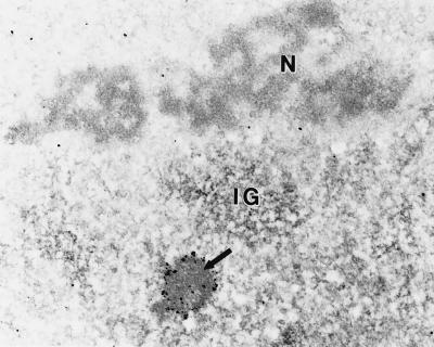
Ribonucleic acid can be detected in the PML-containing nuclear bodies. Immunoelectron microscopy of a nucleus labeled for PML followed by EDTA-regressive staining (36) demonstrates the localization of RNA in subnuclear structures. Using this technique, a PML body (arrow) demonstrates the characteristic decoration of the nuclear body by PML fringing its outer shell as well as positive staining for RNA throughout the entire nuclear body. As expected, RNA was also detected in nucleoli (N) and interchromatin granule (IG) regions. Chromatin surrounding the nucleolus is devoid of staining. (×20,600.)

Characterization of the FITC–UTP foci. When FITC–UTP is co-injected with increasing concentrations of unconjugated UTP, the fluorescent signal is competed away. The photomicrographs represent 0 (a), 50 (b) and 100 (c) mM UTP added along with 1 mM FITC–UTP. HEp-2 cells were nuclearly microinjected with 1 μg/ml of α-amanitin, and after 10 min, the cells were microinjected with FITC–UTP and then processed for microscopic evaluation. Compared with untreated control cells (d), microinjection of α-amanitin (e) inhibits the FITC–UTP labeling of foci and diminishes incorporation into the nucleoplasm but not nucleoli. A corresponding image of PML labeling (f) demonstrates that the structural integrity of the PML body is not affected by α-amanitin. The effect of actinomycin D-mannitol on incorporation of FITC–UTP was examined in cells incubated for 1 h at concentrations of 0 (g), 0.01 (h), and 0.20 (i) μg/ml. Actinomycin D appears to inhibit pol I synthesis at the lower dose and pol I and pol II at the higher dose.
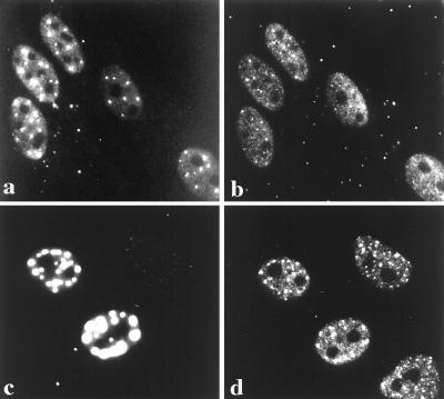
CBP colocalizes with the PML-containing nuclear body. Double labeling of HEp-2 cells demonstrate PML-containing nuclear bodies as detected with the murine monoclonal antibody 5E10 (a). In the same cells, endogenous CBP is distributed in a finely speckled nucleoplasmic pattern as well as on larger nuclear foci, which can only be observed when utilizing the affinity-purified anti-CBP directed against amino acids 634–648 (b). Comparison of the images demonstrates that CBP and PML colocalize to the same nuclear bodies. To confirm this colocalization, cells exogenously expressing the PML-GFP fusion protein (c) were immunostained for CBP (d), which not only colocalized to the PML-containing nuclear body but appeared to label with a stronger signal, suggesting a recruitment of CBP to the PML bodies.
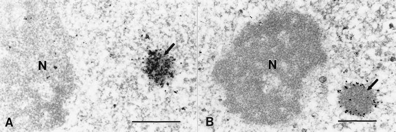
Ultrastructural analysis of CBP and PML localization in HEp2 cells by immunoelectron microscopy. Silver-enhanced Nanogold pre-embedding immunoelectron microscopy (35) was performed utilizing affinity-purified rabbit antibodies to CBP (aa 634–648) and PML (aa 1–14). (A) CBP was localized in a nuclear body (arrow) with homogeneous labeling throughout. (B) PML localization was restricted to the outer shell of the PML-containing nuclear body (arrow). Nucleoli (N) were unlabeled. (Bar = 0.5 μm.)
References
-
- Guiochon-Mantel A, Savouret J, Quignon F, Delabre K, Milgrom E, De The H. Mol Endocrinol. 1995;9:1791–1803. - PubMed
-
- Rowley J D, Golomb H M, Dougherty C. Lancet. 1977;1:549–550. - PubMed
-
- de The H, Chomienne C, Lanotte M, Degos L, Dejean A. Nature. 1990;347:558–561. - PubMed
-
- Kakizuka A, Miller W, Jr, Umesono K, Warrell R, Jr, Frankel S R, Murty V V, Dmitrovsky E, Evans R M. Cell. 1991;66:663–674. - PubMed
Publication types
MeSH terms
Substances
Grants and funding
LinkOut - more resources
Full Text Sources
Other Literature Sources
