

doi: 10.1073/pnas.95.9.5150.
Monosomy of a specific chromosome determines L-sorbose utilization: a novel regulatory mechanism in Candida albicans
Affiliations
- PMID: 9560244
- PMCID: PMC20229
- DOI: 10.1073/pnas.95.9.5150
Item in Clipboard
Monosomy of a specific chromosome determines L-sorbose utilization: a novel regulatory mechanism in Candida albicans
Proc Natl Acad Sci U S A.
.
Abstract
We report the identification of the gene, SOU1, required for L-sorbose assimilation in Candida albicans. The level of the expression of SOU1 is determined by the copy number of chromosome III (also denoted chromosome 5), such that monosomic strains assimilate L-sorbose, whereas disomic strains do not, in spite of the fact that SOU1 is not on this chromosome. We suggest that C. albicans contains a resource of potentially beneficial genes that are activated by changes in chromosome number, and that this elaborate mechanism regulates the utilization of food supplies and possibly other important functions, thus representing a novel general means for regulating gene expression in microbes.
Figures
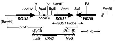
Physical map of the SOU1 region
showing SOU2 and VMA8 flanking genes; the
C-rich 40-bp poly(C) segment; P1, P2, and P3 primers used to generate
PCR fragments; SOU1 probe used in Southern and Northern
blot analysis; and a portion of the pCA7 plasmid used for disrupting
SOU1.
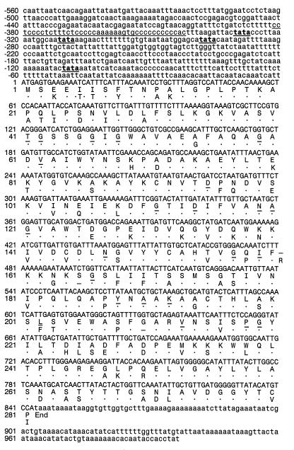
Nucleotide sequence of the SOU1
gene and deduced amino acid sequence of the corresponding Sou1p. The
residues conserved in the short-chain alcohol dehydrogenase family are
underlined. The C-rich domain and the putative TATA elements are
underlined in the 5′ untranslated region. The Sou2p sequence is shown
below the Sou1p sequence, where identical residues are depicted by
dots.
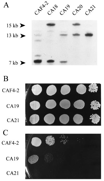
(A) Southern blot analysis of DNA
digestion with BglII and hybridization with the
SOU1 probe of the following strains: CAF4–2, the normal
SOU1/SOU1 strain; CA18, the singly
disrupted
SOU1/sou1-Δ∷hisG-URA3-hisG
strain; CA19, the singly disrupted
SOU1/sou1-Δ∷hisG
strain; CA20, the doubly disrupted
sou1-Δ∷hisG/sou1-Δ∷hisG-URA3-hisG
strain; and CA21, the doubly disrupted
sou1-Δ∷hisG/sou1-Δ∷hisG
strain. (B) Comparative growth of CAF4–2 and CA19 and CA21
on glucose medium (SD plus uridine) for 6 days at 37°C and
(C) on l -sorbose medium (plus uridine) for 14
days at 37°C. Each strain was spotted as serial 1/10 dilutions,
from left to right, starting from an initial concentration of
106 cells per ml.
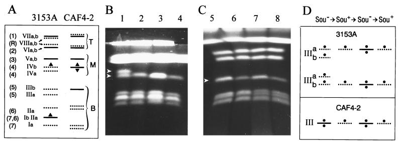
(A) Schematic representation of the
electrophoretic karyotypes from 3153A and CAF4–2 showing the positions
of the SOU1 gene (▴ and ▾).
Orthogonal-field-alternation gel electrophoresis separation of
chromosomes of two typical representative sequential series of strains
derived from the parental strains (B) 3153A and
(C) CAF4–2. Orthogonal-field-alternation gel
electrophoresis. Lanes: 1, 3153A (Sou−); 2, Sor55
(Sou+); 3, Sor55–1 (Sou−); 4, Sor55–1-1
(Sou+); 5, CAF4–2 (Sou−); 6, Sor19
(Sou+); 7, Sor19–1 (Sou−); 8, Sor19–1-1
(Sou+). Conditions of separation in these two
representative gels were selected for the best resolution of either the
smallest or the smallest and middle-sized groups of chromosomes (21).
The gels showing precise separations of the other chromosomes of these
strains are not presented. (D) Schematic representation of
chromosomes III from the Sou+ and Sou− strains
serially derived from 3153A and CAF4–2 showing the hypothetical
negative regulator CSU1 gene (•).
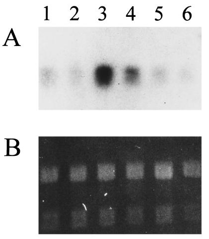
(A) Expression of SOU1
as determined by Northern blot analysis with a SOU1
probe and with the following strains. Lanes: 1 and 2, parental strain
3153A (Sou−); 3, Sor52 (Sou+); 4, Sor53
(Sou+); 5, Sor52–1 (Sou−); and 6, Sor53–1
(Sou−). (B) Relative SOU1 mRNA
levels were estimated by comparing the hybridization signals with the
ethidium bromide fluorescence intensities of the rRNA bands.
References
Publication types
MeSH terms
Substances
Associated data
- Actions
Grants and funding
LinkOut - more resources
Full Text Sources
Other Literature Sources
Molecular Biology Databases
