

CD40 ligand (CD154) stimulation of macrophages to produce HIV-1-suppressive beta-chemokines
- PMID: 9560254
- PMCID: PMC20239
- DOI: 10.1073/pnas.95.9.5205
CD40 ligand (CD154) stimulation of macrophages to produce HIV-1-suppressive beta-chemokines
Abstract
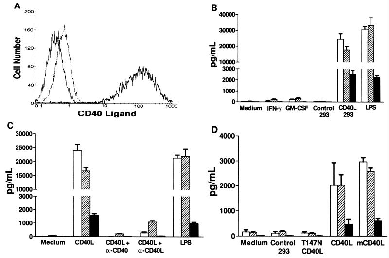
beta-chemokines play an important role in the development of immunologic reactions. Macrophages are major beta-chemokine-producing cells during T-cell directed, delayed-type hypersensitivity reactions in tissues, and have been reported to be important producers of beta-chemokines in the lymph nodes of HIV-1-infected individuals. However, the physiological signals responsible for inducing macrophages to produce beta-chemokines have not been established. Two soluble T cell products, interferon-gamma and granulocyte-macrophage colony stimulating factor, were added to cultured macrophages, but failed to stimulate the production of macrophage inflammatory protein-1alpha and -1beta; regulated upon activation, normal T cell expressed and secreted (RANTES); or monocyte chemoattractant protein-1. Instead, direct cell-cell contact between macrophages and cells engineered to express CD40L (also known as CD154) resulted in the production of large amounts of macrophage inflammatory protein-1alpha and -1beta, and RANTES (all ligands for CCR5), and monocyte chemoattractant protein-1 (a ligand for CCR2). Supernatants from CD40L-stimulated macrophages protected CD4(+) T cells from infection by a nonsyncytium-inducing strain of HIV-1 (which uses CCR5 as a coreceptor). These results have implications for granulomatous diseases, and conditions such as atherosclerosis and multiple sclerosis, where CD40L-bearing cells have been found in the macrophage-rich lesions where beta-chemokines are being produced. Overall, these findings define a pathway linking the specific recognition of antigen by T cells to the production of beta-chemokines by macrophages. This pathway may play a role in anti-HIV-1 immunity and the development of immunologic reactions or lesions.
Figures


Similar articles
-
Regulation of human immunodeficiency virus type 1 infection, beta-chemokine production, and CCR5 expression in CD40L-stimulated macrophages: immune control of viral entry.J Virol. 2001 May;75(9):4308-20. doi: 10.1128/JVI.75.9.4308-4320.2001. J Virol. 2001. PMID: 11287580 Free PMC article.
-
Differential effects of CD40 ligand/trimer stimulation on the ability of dendritic cells to replicate and transmit HIV infection: evidence for CC-chemokine-dependent and -independent mechanisms.J Immunol. 1999 Mar 15;162(6):3711-7. J Immunol. 1999. PMID: 10092834
-
Soluble CD40 ligand induces beta-chemokine production by macrophages and resistance to HIV-1 entry.Cytokine. 2000 Oct;12(10):1489-95. doi: 10.1006/cyto.1999.0594. Cytokine. 2000. PMID: 11023663
-
The emerging role of CD40 ligand in HIV infection.J Leukoc Biol. 2000 Sep;68(3):373-82. J Leukoc Biol. 2000. PMID: 10985254 Review.
-
Cytokines and HIV-1: interactions and clinical implications.Antivir Chem Chemother. 2001 May;12(3):133-50. doi: 10.1177/095632020101200301. Antivir Chem Chemother. 2001. PMID: 12959322 Review.
Cited by
-
Fractalkine (CX3CL1) and brain inflammation: Implications for HIV-1-associated dementia.J Neurovirol. 2002 Dec;8(6):585-98. doi: 10.1080/13550280290100950. J Neurovirol. 2002. PMID: 12476352 Review.
-
The macrophage in HIV-1 infection: from activation to deactivation?Retrovirology. 2010 Apr 9;7:33. doi: 10.1186/1742-4690-7-33. Retrovirology. 2010. PMID: 20380696 Free PMC article. Review.
-
Induction of rapid and extensive beta-chemokine synthesis in macrophages by human immunodeficiency virus type 1 and gp120, independently of their coreceptor phenotype.J Virol. 2001 Nov;75(22):10738-45. doi: 10.1128/JVI.75.22.10738-10745.2001. J Virol. 2001. PMID: 11602715 Free PMC article.
-
Levels of soluble CD40 ligand and P-Selectin in nonalcoholic fatty liver disease.Dig Dis Sci. 2010 Apr;55(4):1128-34. doi: 10.1007/s10620-009-0817-1. Epub 2009 May 14. Dig Dis Sci. 2010. PMID: 19440836
-
Activation of tolerogenic dendritic cells in the tumor draining lymph nodes by CD8+ T cells engineered to express CD40 ligand.J Immunol. 2010 Apr 1;184(7):3394-400. doi: 10.4049/jimmunol.0903111. Epub 2010 Mar 3. J Immunol. 2010. PMID: 20200275 Free PMC article.
References
-
- Taub D D, Oppenheim J J. Therapeutic Immunol. 1994;1:229–246. - PubMed
-
- Schall T J, Bacon K B. Curr Opin Immunol. 1994;6:865–873. - PubMed
-
- Murphy P M. Annu Rev Immunol. 1994;12:593–633. - PubMed
-
- Sherry B, Horii Y, Manogue K R, Widmer U, Cerami A. Cytokines. 1992;4:117–130. - PubMed
-
- Schall T J, Jongstra J, Dyer B J, Jorgensen J, Clayberger C, Davis M M, Krensky A M. J Immunol. 1988;141:1018–1025. - PubMed
Publication types
MeSH terms
Substances
Grants and funding
LinkOut - more resources
Full Text Sources
Other Literature Sources
Molecular Biology Databases
Research Materials
