. 1998 Apr 28;95(9):5251-6.
doi: 10.1073/pnas.95.9.5251.

Modulation of ventricular function through gene transfer in vivo

Affiliations

Modulation of ventricular function through gene transfer in vivo

R J Hajjar et al. Proc Natl Acad Sci U S A. .

Abstract

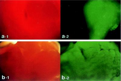
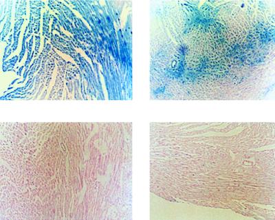
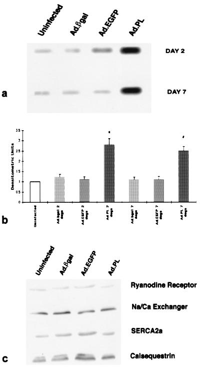
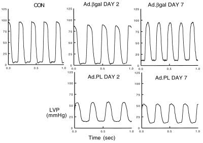
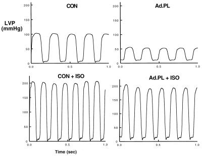
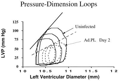
We used a catheter-based technique to achieve generalized cardiac gene transfer in vivo and to alter cardiac function by overexpressing phospholamban (PL) which regulates the activity of the sarcoplasmic reticulum Ca2+ ATPase (SERCA2a). By using this approach, rat hearts were transduced in vivo with 5 x 10(9) pfu of recombinant adenoviral vectors carrying cDNA for either PL, beta-galactosidase (beta-gal), or modified green fluorescent protein (EGFP). Western blot analysis of ventricles obtained from rats transduced by Ad.PL showed a 2.8-fold increase in PL compared with hearts transduced by Ad.betagal. Two days after infection, rat hearts transduced with Ad.PL had lower peak left ventricular pressure (58.3 +/- 12.9 mmHg, n = 8) compared with uninfected hearts (92.5 +/- 3.5 mmHg, n = 6) or hearts infected with Ad.betagal (92.6 +/- 5.9 mmHg, n = 6). Both peak rate of pressure rise and pressure fall (+3, 210 +/- 298 mmHg/s, -2, 117 +/- 178 mmHg/s, n = 8) were decreased in hearts overexpressing PL compared with uninfected hearts (+5, 225 +/- 136 mmHg/s, -3, 805 +/- 97 mmHg/s, n = 6) or hearts infected with Ad.betagal (+5, 108 +/- 167 mmHg/s, -3, 765 +/- 121 mmHg/s, n = 6). The time constant of left ventricular relaxation increased significantly in hearts overexpressing PL (33.4 +/- 3.2 ms, n = 8) compared with uninfected hearts (18.5 +/- 1.0 ms, n = 6) or hearts infected with Ad.betagal (20.8 +/- 2.1 ms, n = 6). These differences in ventricular function were maintained 7 days after infection. These studies open the prospect of using somatic gene transfer to modulate overall cardiac function in vivo for either experimental or therapeutic applications.


Figures

Figure 1
Figure 1
Rat hearts were transduced with Ad.EGFP by using either the catheter-based technique (b) or direct injection into the left ventricular wall (a). Forty-eight hours following delivery of adenovirus encoding for EGFP, the left ventricles of the hearts were removed and visualized with white light (a-1 and b-1) and at 510 nm with single excitation peak at 490 nm of blue light (a-2 and b-2). As shown in b-2, the expression pattern observed after catheter delivery is grossly homogeneous. In contrast, the expression pattern is localized after direct injection as shown in a-2. Of note, with the direct injection, the surrounding tissue exhibits no background fluorescence (a-2).
Figure 2
Figure 2
Expression of β-gal in left ventricular sections 2 days following infection with Ad.βgal and Ad.PL. (Upper) Photomicrographs of two left ventricular sections stained for β-gal 2 days following infection with Ad.βgal. These sections show the variability of β-gal expression within the same heart with the catheter-based method of gene delivery. (Lower) Photomicrographs of two left ventricular sections stained for β-gal 2 days following infection with Ad.PL. No β-gal expression is observed.
Figure 3
Figure 3
(a) Immunoblots of PL from crude membranes of left ventricles from control uninfected rats or rats 2 and 7 days after infection with 5 × 10⁹ pfu of either Ad.βgal, Ad.EGFP, or Ad.RSV.PL. (b) Protein levels of PL in preparations from uninfected hearts (n = 8), preparations of hearts infected with Ad.βgal at day 2 (n = 6), preparations of hearts infected with Ad.EGFP at day 2 (n = 4), preparations of hearts infected with Ad.PL at day 2 (n = 8), preparations of hearts infected with Ad.βgal at day 7 (n = 6), preparations of hearts infected with Ad.EGFP at day 7 (n = 4), and preparations of hearts infected with Ad.PL at day 7 (n = 8). ∗, P < 0.05 compared with uninfected, Ad.βgal at 2 days, and Ad.EGFP at 2 days. #, P < 0.05 compared with Ad.βgal at 2 days, and Ad.EGFP at 7 days. There were no significant differences between the PL protein levels in the uninfected group and Ad.βgal or Ad.EGFP (P > 0.2). (c) Immunoblots with mAbs to the of ryanodine receptor, Na/Ca exchanger, SERCA2a, and calsequestrin from crude membrane of left ventricles after 2 days of infection with 5 × 10⁹ pfu of either Ad.βgal, Ad.EGFP, or Ad.RSV.PL.
Figure 4
Figure 4
Left ventricular pressure measurements from rats that were either uninfected (CON) or infected with Ad.βgal or Ad.PL 2 and 7 days after infection as indicated. Hearts infected with Ad.PL had a decrease in systolic pressure, elevation of diastolic pressure, and prolongation of the relaxation phase.
Figure 5
Figure 5
Left ventricular pressure measurements during infusion of 0.1 μg/kg/min of isoproterenol in an uninfected rat heart and rat hearts infected with Ad.PL (5 × 10⁹ pfu, day 2).
Figure 6
Figure 6
Left ventricular pressure vs. left ventricular dimension detected by piezoelectric crystals in a control uninfected heart and in a heart infected with Ad.PL (5 × 10⁹ pfu, day 2) under different loads obtained by clamping the inferior vena cava.

References

    1. Barry W H, Bridge J H B. Circulation. 1993;87:1806–1815. - PubMed
    1. Gwathmey J K, Bentivegna L A, Ransil B J, Grossman W, Morgan J P. Cardiovasc Res. 1993;27:199–203. - PubMed
    1. Koss K L, Kranias E G. Circulation Res. 1996;79:1059–1063. - PubMed
    1. Arai M, Matsui H, Periasamy M. Circ Res. 1994;74:555–564. - PubMed
    1. Hajjar R J, Kang J X, Gwathmey J K, Rosenzweig A. Circulation. 1997;95:423–429. - PubMed

Publication types

LinkOut - more resources