. 1998 Apr 28;95(9):5372-7.
doi: 10.1073/pnas.95.9.5372.

Targeted inactivation of Npt2 in mice leads to severe renal phosphate wasting, hypercalciuria, and skeletal abnormalities

Affiliations

Targeted inactivation of Npt2 in mice leads to severe renal phosphate wasting, hypercalciuria, and skeletal abnormalities

L Beck et al. Proc Natl Acad Sci U S A. .

Abstract

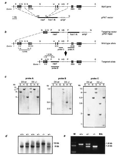
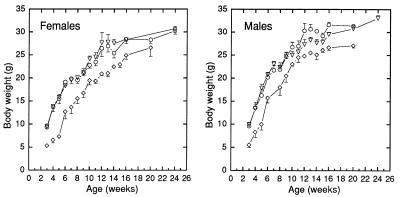
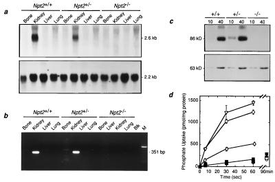
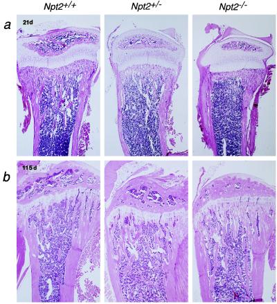
Npt2 encodes a renal-specific, brush-border membrane Na+-phosphate (Pi) cotransporter that is expressed in the proximal tubule where the bulk of filtered Pi is reabsorbed. Mice deficient in the Npt2 gene were generated by targeted mutagenesis to define the role of Npt2 in the overall maintenance of Pi homeostasis, determine its impact on skeletal development, and clarify its relationship to autosomal disorders of renal Pi reabsorption in humans. Homozygous mutants (Npt2(-/-)) exhibit increased urinary Pi excretion, hypophosphatemia, an appropriate elevation in the serum concentration of 1,25-dihydroxyvitamin D with attendant hypercalcemia, hypercalciuria and decreased serum parathyroid hormone levels, and increased serum alkaline phosphatase activity. These biochemical features are typical of patients with hereditary hypophosphatemic rickets with hypercalciuria (HHRH), a Mendelian disorder of renal Pi reabsorption. However, unlike HHRH patients, Npt2(-/-) mice do not have rickets or osteomalacia. At weaning, Npt2(-/-) mice have poorly developed trabecular bone and retarded secondary ossification, but, with increasing age, there is a dramatic reversal and eventual overcompensation of the skeletal phenotype. Our findings demonstrate that Npt2 is a major regulator of Pi homeostasis and necessary for normal skeletal development.


Figures

Figure 1
Figure 1
Targeted disruption of the murine Npt2 gene. (a) Production of the pPNT–Npt2 targeting vector. (Upper) Schematic representation of the murine Npt2 gene, with exons numbered and denoted as grey boxes. (Lower) pPNT vector containing the neor and hsv-tk genes. The broken lines indicate sites where the Npt2 homologous arms were inserted. Relevant restriction enzyme sites are abbreviated as follows: E, EcoRI; H, HindIII; N, NotI; P, PstI; S, SstI; X, XhoI. (b) Targeting of Npt2 by homologous recombination. The top line represents the incoming pPNT–Npt2 targeting vector, the middle line the normal Npt2 allele, and the bottom line the targeted allele. The location of the probes used in Southern blot analysis are indicated. Probe A, 1.5-kb fragment external to the targeting vector; probe B, corresponding to the neor gene; probe C, a 0.8-kb fragment used as an internal probe. (c) Southern blot analysis of targeted ES cell clones. Genomic DNA (5 μg) derived from untransfected ES cells (D3 wt) or from targeted clones (D3 +/−) was digested with EcoRI (E), HindIII (H), and SstI (S), Southern blotted, and hybridized with probes A, B, and C, as shown. The sizes of genomic DNA fragments expected from the normal and disrupted alleles are indicated. (d) Genotyping of 3-week-old offspring from heterozygous matings by Southern blot analysis. Probe A was hybridized to EcoRI-restricted tail genomic DNA. Wild-type (+/+) and heterozygous (+/−) animals exhibited an 8-kb band that is absent in homozygous mutant (−/−) mice. Disruption of the Npt2 allele produced a 12-kb band. (e) PCR analysis of mouse tail DNA from wild-type (+/+), heterozygous (+/−), and homozygous mutant (−/−) mice. Positions of the primers used for the PCR and the expected sizes of amplified fragments are indicated above the corresponding alleles in b. M, size markers; Blk, negative control.
Figure 2
Figure 2
Body weight of Npt2+/+, Npt2+/−, and Npt2−/− mice as a function of age. Normal mice and their mutant littermates were weighed between 3 weeks and 6 months of age. Each point represents mean ± SD derived from 4 to 30 measurements. Npt2+/+ (○), Npt2+/− (▿), and Npt2−/− (⋄) mice.
Figure 3
Figure 3
Npt2 gene expression. (a) Northern blot analysis of total RNA from bone, kidney, liver, and lung of Npt2+/+, Npt2+/− and Npt2−/− mice. Total RNA (15 μg) was blotted to a nylon membrane and hybridized sequentially with 32P-labeled rat Npt2 cDNA (Upper) and 18S rRNA oligonucleotide (Lower) probes as described. (b) RT-PCR of total RNA from Npt2+/+, Npt2+/−, and Npt2−/− mice. Total RNA (5 μg) from bone, kidney, liver, and lung was reverse transcribed and PCR amplified as described. An aliquot of each PCR was electrophoresed on 1.5% agarose gels and visualized with ethidium bromide. M, size markers, Blk, negative control. (c) Western blot analysis of renal BBM proteins from Npt2+/+, Npt2+/, and Npt2−/− mice. The BBMs were solubilized and subjected to SDS/PAGE, transferred to nitrocellulose, and the blots probed sequentially with (Upper) a rabbit polyclonal anti-rat Npt2 antibody and (Lower) a mAb raised against the α-subunit of rat meprin. Molecular masses and quantity of protein (μg) applied to each lane are indicated. (d) Pi uptake in renal BBM vesicles from Npt2+/+, Npt2+/, and Npt2−/− mice. Pi uptake (100 μM) was measured at times indicated in presence of KCl (filled symbols) and NaCl (open symbols) in BBM vesicles derived from Npt2+/+ (circles), Npt2+/ (triangles), and Npt2−/− (diamond) mice as described. Each point represents mean ± SEM of quadruplicate determinations.
Figure 4
Figure 4
Histological appearance of bone from Npt2+/+, Npt2+/−, and Npt2−/− mice. Longitudinal tibial sections were prepared from 21- (a) and 115-day-old (b) mice as described and stained with hematoxylin/eosin.

References

    1. Mizgala C L, Quamme G A. Physiol Rev. 1985;65:431–466. - PubMed
    1. Murer H, Werner A, Reshkin S, Wuarin R, Biber J. Am J Physiol. 1991;260:C885–C899. - PubMed
    1. Berndt T J, Knox F G. In: The Kidney: Physiology and Pathophysiology. Seldin D W, Giebisch G, editors. New York: Raven; 1992. pp. 2511–2532.
    1. Walker J J, Yan T S, Quamme G A. Am J Physiol. 1987;252:F226–F231. - PubMed
    1. Werner A, Moore M L, Mantei N, Biber J, Semenza G, Murer H. Proc Natl Acad Sci USA. 1991;88:9608–9612. - PMC - PubMed

Publication types

MeSH terms