. 1998 Apr 28;95(9):5401-6.
doi: 10.1073/pnas.95.9.5401.

Association of the Arabidopsis CTR1 Raf-like kinase with the ETR1 and ERS ethylene receptors

Affiliations

Association of the Arabidopsis CTR1 Raf-like kinase with the ETR1 and ERS ethylene receptors

K L Clark et al. Proc Natl Acad Sci U S A. .

Erratum in

  • Proc Natl Acad Sci U S A 1998 Jul 21;95(15):9060

Abstract

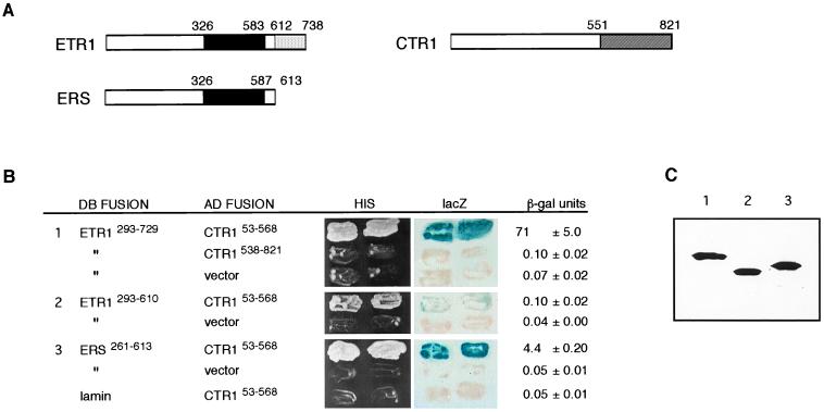
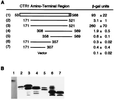
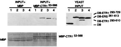
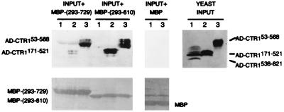
In Arabidopsis thaliana, signal transduction of the hormone ethylene involves at least two receptors, ETR1 and ERS, both of which are members of the two-component histidine protein kinase family that is prevalent in prokaryotes. The pathway also contains a negative regulator of ethylene responses, CTR1, which closely resembles members of the Raf protein kinase family. CTR1 is thought to act at or downstream of ETR1 and ERS based on double mutant analysis; however, the signaling mechanisms leading from ethylene perception to the regulation of CTR1 are unknown. By using the yeast two-hybrid assay, we detected a specific interaction between the CTR1 amino-terminal domain and the predicted histidine kinase domain of ETR1 and ERS. We subsequently verified these interactions by using an in vitro protein association assay(s). In addition, we determined that the amino-terminal domain of CTR1 can associate with the predicted receiver domain of ETR1 in vitro. Based on deletion analysis, the portion of CTR1 that interacts with ETR1 roughly aligns with the regulatory region of Raf kinases. These physical associations support the genetic evidence that CTR1 acts in the pathway of ETR1 and ERS and suggest that these interactions could be involved in the regulation of CTR1 activity.


Figures

Figure 1
Figure 1
Interaction of ETR1 and ERS proteins with the CTR1 protein in the yeast two-hybrid assay. (A) Schematic structures of ETR1, ERS, and CTR1 proteins. Domains are approximated based on sequence homology with two-component proteins and Raf kinases, respectively; the solid black region in ETR1 and ERS indicates the histidine kinase domain, the light-shaded region in ETR1 indicates the receiver domain, and the shaded region of CTR1 indicates the serine/threonine kinase domain. (B) Two-hybrid constructs and results. Regions of ETR1, ERS, and CTR1 that were fused to the DB or AD are indicated by residue numbers. The vector is pGAD424, which produces AD alone. Human lamin was used as a nonspecific control. HIS shows growth of transformants on medium lacking histidine. lacZ shows the X-Gal filter assay of the same transformants (grown in the presence of histidine) after 1 hr at room temperature. β-gal units gives β-galactosidase activity in modified Miller units (22). Three transformants of each were measured, and the average ± the SD is presented. (C) Immunoblot analysis of the DB fusion proteins present in yeast double transformants. The same transformants that were tested in the above assays were analyzed for relative levels of DB fusion proteins. We tested two transformants of each and observed similar levels for each pair; one representative of each is shown. Immunoblot analysis of the AD fusions is shown in Fig. 4.
Figure 2
Figure 2
Localization of the region of CTR1 that associates with ETR1. (A) Results of the yeast two-hybrid assay. β-Galactosidase activity [in modified Miller units (22)] was measured in yeast transformants expressing DB–ETR1293–729 plus each of the indicated AD–CTR1 fusion proteins. Fusions 1 and 2 were expressed in plasmid pGAD424; fusions 3–7 were expressed in plasmid pACTII (which has a stronger promoter than pGAD424). Three transformants of each were tested, and the average ± the SD is presented. The background level of activity for transformants carrying the pACTII vector is given. (B) Immunoblot analysis of the levels of AD fusion proteins in the transformants using antibody to Gal4 (AD). Two of each of the transformants tested for β-galactosidase activity were examined and were found to be similar; a representative of each is shown. The exposure for construct 1 was approximately four times longer than for the other constructs.
Figure 3
Figure 3
In vitro association of purified MBP–CTR153–568 with yeast DB–ETR1 and DB–ERS fusions in yeast cell extracts. Bacterially expressed MBP or MBP–CTR153–568 protein was attached to amylose-containing beads, the beads were mixed with yeast extracts 1 through 4, and then the bead-associated proteins were subjected to immunoblot analysis with antibody to LexA (DB). YEAST INPUT is an immunoblot analysis of the yeast extracts before treatment with the beads and indicates the relative amounts of yeast DB fusion proteins present in each extract. The input yeast extracts were: (lane 1) DB alone, (lane 2) DB–ETR1293–729, (lane 3) DB–ERS261–613, and (lane 4) DB–ETR1293–610. For the samples labeled INPUT + MBP, each indicated yeast extract was mixed with MBP attached to beads. For the samples labeled INPUT + MBP–CTR153–568, each indicated yeast extract was mixed with the MBP–CTR153–568 fusion protein attached to beads. The lower panel shows the total protein on the immunoblot filter as detected by Ponceau S.
Figure 4
Figure 4
In vitro association of purified MBP–ETR1 fusions with yeast AD–CTR1 fusions in yeast cell extracts. Bacterially expressed MBP or the indicated MBP fusion was attached to amylose-containing beads, the beads were mixed with yeast extracts 1 through 3, and then the bead-associated proteins were subjected to immunoblot analysis with antibody to Gal4 (AD). YEAST INPUT is an immunoblot analysis of the yeast extracts before treatment with the beads and indicates the relative amounts of yeast AD fusion proteins present in each extract. The input yeast extracts were: (lane 1) AD–CTR1538–821, (lane 2) AD–CTR1171–521, and (lane 3) AD–CTR153–568. For the samples labeled INPUT + MBP-(293–729), each indicated yeast extract was mixed with the MBP–ETR1293–729 fusion protein attached to beads. For the samples labeled INPUT + MBP-(293–610), the indicated yeast extracts were mixed with the MBP–ETR1293–610 fusion protein attached to beads. For the samples labeled INPUT + MBP, yeast extracts were mixed with MBP attached to beads. The lower panels show the total protein present on the immunoblot filters as detected by Ponceau S. MBP-(293–729) is the MBP–ETR1293–729 fusion, and MBP-(293–610) is the MBP–ETR1293–610 fusion.
Figure 5
Figure 5
In vitro association of radiolabeled CTR1 polypeptides with purified MBP fusions: (i) MBP alone, (ii) MBP–ETR1293–610, (iii) MBP–ETR1604–738, and (iv) MBP-CKI1981–1122. (A) Autoradiograms showing association of the CTR1 amino-terminal domain with MBP fusions 1–4. Bacterially expressed MBP or MBP fusion was attached to amylose-containing beads, and the beads were mixed with 5 or 25 μl of in vitro-translated, radiolabeled CTR1 amino-terminal domain (residues 53–568) (IVT). The bead-associated proteins were separated on SDS/PAGE gels, and the radiolabeled CTR153–568 was visualized by autoradiography. Lane IVT contains 0.1 μl of unassociated radiolabeled CTR153–568. (B) Autoradiogram showing association of the CTR1 kinase domain with MBP fusions 1–3. Bacterially expressed MBP or MBP fusion was attached to amylose-containing beads, and the beads were mixed with 5 μl of IVT. IVT in this case is the radiolabeled in vitro-translated CTR1 kinase domain (residues 538–821). The bead-associated proteins were subjected to SDS/PAGE, and radiolabeled CTR1538–821 was visualized by autoradiography. Lane IVT contains 0.1 μl of unassociated radiolabeled CTR1538–821. The length of exposure is twice that shown in Fig. 5A. (C) Relative amounts of MBP fusions 1–4 used in Fig. 5 A and B separated on SDS/PAGE gels and stained with Coomassie blue.

References

    1. Abeles F B, Morgan P W, Saltveit M E., Jr . Ethylene in Plant Biology. 2nd Ed. New York: Academic; 1992.
    1. Chang C, Kwok S F, Bleecker A B, Meyerowitz E M. Science. 1993;262:539–544. - PubMed
    1. Hua J, Chang C, Sun Q, Meyerowitz E M. Science. 1995;269:1712–1714. - PubMed
    1. Kieber J J, Rothenberg M, Roman G, Feldmann K A, Ecker J R. Cell. 1993;72:427–441. - PubMed
    1. Chao Q, Rothenberg M, Solano R, Roman G, Terzaghi W, Ecker J R. Cell. 1997;89:1133–1144. - PubMed

Publication types