Review
. 1998 Apr;11(2):366-81.
doi: 10.1128/CMR.11.2.366.

Zoonotic filariasis

Affiliations
Review

Zoonotic filariasis

T C Orihel et al. Clin Microbiol Rev. 1998 Apr.

Abstract

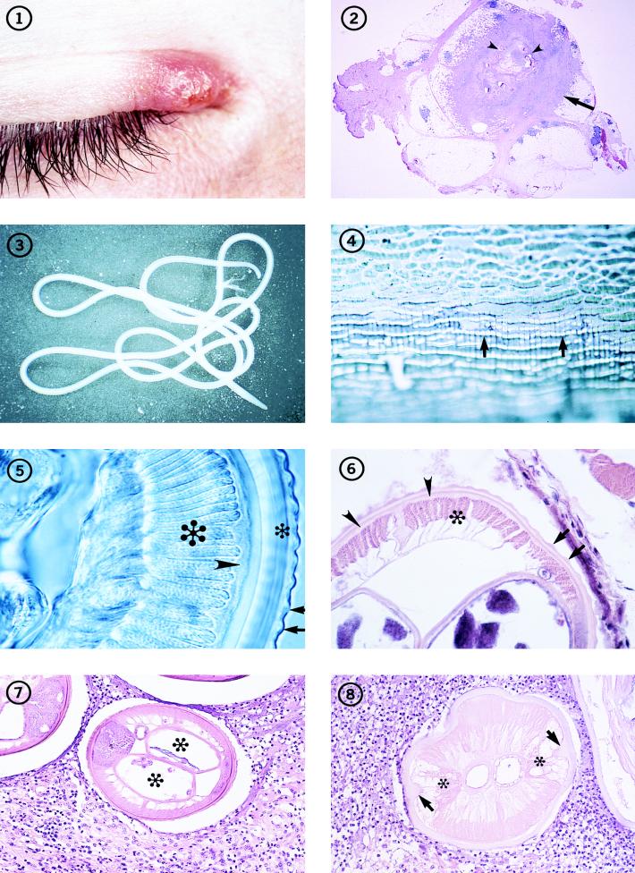
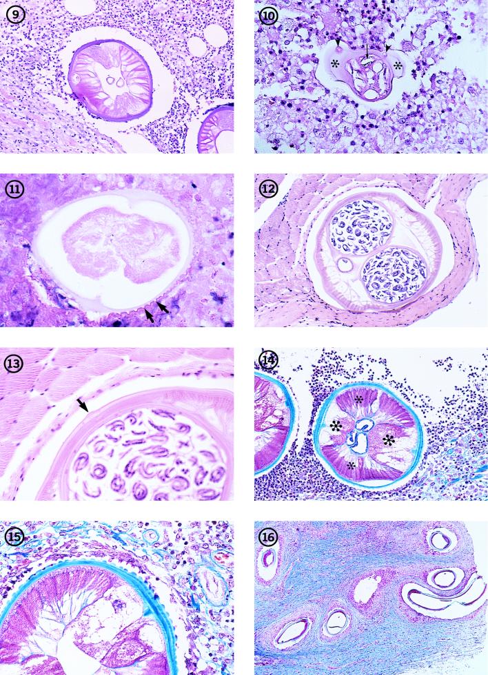
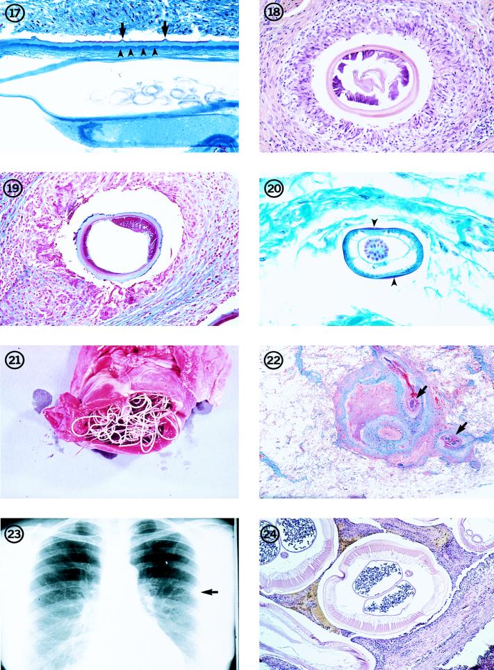
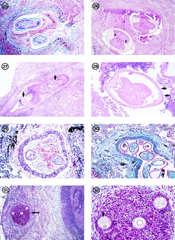
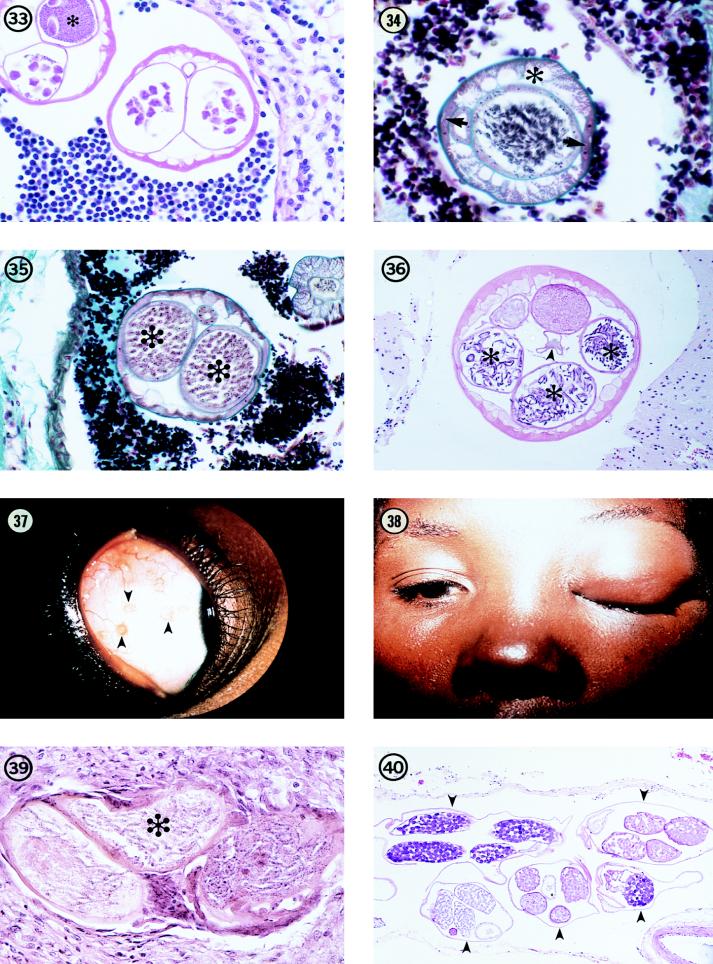
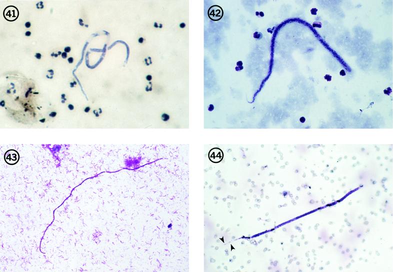
Filariae of animals, especially those of mammals, often infect humans and typically produce cryptic infections. These "zoonotic" infections have been reported from virtually all parts of the world including temperate zones. Infections may be symptomatic or not, and the parasites are found in surgical tissue biopsy specimens or, more rarely, are removed intact from superficial sites such as the orbit or conjuctivae. Typically, these worms tend to occupy tissue sites similar to those occupied in the natural animal host, with the exception of the eyes. Many kinds of filariae have been isolated from humans, including species of Dirofilaria, Brugia, Onchocerca, Dipetalonema, Loaina and Meningonema. Worms have been found in subcutaneous tissues, the heart and lungs, lymphatics, the eye, and the central nervous system. Specific identification of these filariae is based on their morphological features in histologic sections. Unfortunately, some of these worms cannot be identified even at the generic level. There are other species of filariae, presumed to be zoonotic, which produce patent infections in humans but are poorly and incompletely known. These include Microfilaria semiclarum and Microfilaria bolivarensis. It is probable that almost any filaria parasitizing animals can, under proper circumstances, infect humans and undergo some degree of development. Undoubtedly, additional species of filariae will continue to be isolated from humans in the future.


Figures

FIG. 1-8
FIG. 1-8
Individual with an inflammatory lesion at the inner canthus of the eye from which an immature adult D. tenuis worm was removed. Fig. 2 Subcutaneous nodule (arrow) removed from a patient in Florida, containing several sections of a D. tenuis worm (arrowheads). Hematoxylin and eosin stain; magnification, ×4.5. Fig. 3 An immature adult female Dirofilaria species that measured more than 12 cm in length, removed intact from human subcutaneous tissues. Fig. 4 The surface of the cuticle of D. repens, illustrating the beaded appearance of the longitudinal ridges (arrows). The worm was preserved and mounted in glycerin. Magnification, ×182. Fig. 5 Section through an adult D. repens worm, illustrating the appearance of the longitudinal ridges (arrows) in transverse section, as well as the thick, multilayered cuticle (small asterisk), hypodermis (arrowhead), and muscle cells (large asterisk). The worm is unstained but cleared in glycerin. Magnification, ×391. Fig. 6 Portion of a transverse section of D. tenuis, showing the longitudinal ridges (arrows) on the surface of the multilayered cuticle, the thin hypodermis (arrowheads), and the coelomyarian muscles (asterisk). Hematoxylin and eosin stain; magnification, ×227. Fig. 7 Transverse section through a female D. tenuis worm removed from human subcutaneous tissues, illustrating its characteristic morphologic features. The paired uterine tubes (asterisks) contain unsegmented eggs. Hematoxylin and eosin stain; magnification, ×121. Fig. 8 Dead, degenerating female Dirofilaria worm, with a thickened cuticle, prominent lateral internal cuticular ridges (arrows), and frothy, vacuolated lateral chords (asterisks). In the pseudocelom, the tube to the left is the vagina; adjacent to it is the intestine. This section is cut in the anterior one-fifth of the worm. Hematoxylin and eosin stain; magnification, ×164.
FIG. 9-16
FIG. 9-16
Transverse section through a dead, degenerating male D. tenuis worm in human subcutaneous tissues. Note the two tubes in the body cavity (pseudocelom). One is the intestine; the other is the reproductive tube. Hematoxylin and eosin stain; magnification, ×121. Fig. 10 Section through the tail of a dead, male D. tenuis worm. Note the large caudal alae (asterisks) in which caudal papillae are embedded (arrowheads). Note also the presence (in transverse section) of one of the cuticularized spicules (arrow). Hematoxylin and eosin stain; magnification, ×136. Fig. 11 D. ursi-like worm in an advanced state of degeneration in human subcutaneous tissues. Note especially the sharply crested, longitudinal ridges on the surface of the cuticle (arrows). Hematoxylin and eosin stain; magnification, ×573. Fig. 12 Transverse section through a gravid, female D. striata worm from the tissues of a bobcat (Lynx rufus), illustrating its typical dirofilariid morphologic features and especially the absence of conspicuous longitudinal ridges on the surface of the cuticle. Hematoxylin and eosin stain; magnification, ×82. Fig. 13 Higher magnification of the worm in Fig. 12. The relatively inconspicuous lateral ala on the surface of the cuticle is clearly evident (arrow). Hematoxylin and eosin stain; magnification, ×227. Fig. 14 Dead female D. repens worm removed from human subcutaneous tissues. Note the conspicuous longitudinal ridges, the enlarged, frothy lateral chords (large asterisks), and the strong musculature (small asterisks), which retains some of its normal architecture. Trichrome stain; magnification, ×64. Fig. 15 Higher magnification of the worm in Fig. 14. Note the multilayered structure of the cuticle and the irregular spacing of the longitudinal ridges on its surface. Trichrome stain; magnification, ×191. Fig. 16 Section of nodule removed from a patient in Illinois and containing a zoonotic Onchocerca worm. The highly coiled nature of the worm and the dense connective tissue surrounding the worm are evident. Trichrome stain; magnification, ×191. (From the case reported in reference .)
FIG. 17-24
FIG. 17-24
Longitudinal section through a female O. cervicalis worm from a naturally infected horse, illustrating the morphologic features of the cuticle, including the outer circular ridges (arrows) and the inner striae (arrowheads). There are four striae per ridge. Trichrome stain; magnification, ×209. Fig. 18 Transverse section through a female Onchocerca worm removed from a human in Japan. The variable thickness of the cuticle is evident, as is the weak musculature. Two uterine reproductive tubes are present in the body cavity. Hematoxylin and eosin stain; magnification, ×114. (From the case presented in reference .) Fig. 19 Transverse section of the female Onchocerca worm illustrated in Fig. 16. Note the unevenly thick cuticle and the low, weak, poorly developed musculature. Trichrome stain; magnification, ×114. (From the case reported in reference .) Figure 20 Section through a male O. gutturosa worm from a naturally infected cow, illustrating the thin cuticle which thickens laterally (arrowheads), small number of muscle cells, and large testis, which fills the body cavity. The intestine is the small, collapsed tube adjacent to the testis. Trichrome stain; magnification, ×328. Fig. 21 Adult D. immitis in the right ventricle of the heart of its natural host, the domestic dog. Reprinted from reference with permission of the publisher. Fig. 22A coin lesion in a human lung, involving a small pulmonary artery. Sections of an immature D. immitis worm (arrows) can be seen in the obstructed vessel. Hematoxylin and eosin stain; magnification, ×3.6. Fig. 23 Chest X ray showing a typical coin lesion (arrow) in a human lung as a result of a D. immitis infection. Fig. 24 Transverse section of an adult female D. immitis worm, illustrating the characteristic morphologic features of this parasite. Hematoxylin and eosin stain; magnification, ×45.
FIG. 25-32
FIG. 25-32
Dead D. immitis worm in an occluded small pulmonary artery in a human lung. The parasite can be identified by its morphologic features, including its size and the thick but smooth cuticle with prominent lateral, internal cuticular ridges. Hematoxylin and eosin stain; magnification, ×45. Fig. 26 Dead, degenerating, female D. immitis worm in a small pulmonary nodule. Note the thick, swollen cuticle with prominent internal cuticular ridges (arrowheads). The three tubes in the pseudocelom (two reproductive tubes and an intestine) indicate that it is a female worm. Hematoxylin and eosin stain; magnification, ×91. Fig. 27 Low-power view of sections of D. repens (arrows) in an obstructed pulmonary artery of an individual in Italy. Hematoxylin and eosin stain; magnification, ×11.4. Reprinted from reference with permission of the publisher. Fig. 28 Higher magnification of one of the sections of the worm in Fig. 27. The parasite is in an advanced state of degeneration as indicated by the swollen cuticle and degeneration of the soft tissues of the worm. However, longitudinal ridges can be seen on the cortical surface of the cuticle (arrows). Hematoxylin and eosin stain; magnification, ×91. Reprinted from reference with permission of the publisher. Fig. 29 Section through a small pulmonary artery removed from an individual who had lived in India, showing two transverse sections through a Brugia-like worm. Hematoxylin and eosin stain; magnification, ×91. Reprinted from reference with permission of the publisher. Fig. 30 W. bancrofti-like worm lying in a small pulmonary artery of a soldier who had served in Singapore. Trichrome stain; magnification, ×45. (From the case presented in reference .) Fig. 31 Section through a peripheral lymph node from the upper arm of a patient in Ohio. Three sections of a small Brugia worm are present in an occluded lymph channel (arrow) lying inside the nodular capsule. The medullary portion of the lymph node is visible to the right of the nodule. Hematoxylin and eosin stain; magnification, ×59. Fig. 32 Higher magnification of the worm in Fig. 31. Two sections through the anterior end at the level of the esophagus and ovejector (right and left sections) and one section through the region of the intestine and vagina uterina (middle section) are evident. The thin body wall, triradiate esophagus (arrow), few muscle cells per quadrant, and the muscular vagina vera and uterina are evident. Hematoxylin and eosin stain; magnification, ×237.
FIG. 33-40
FIG. 33-40
Two sections of a gravid female Brugia from a zoonotic infection in Peru. In both sections, the paired uterine tubes are evident and contain developing eggs or, in the upper left-hand section, sperm (asterisk), indicating that a male worm was present and that mating had occurred. Hematoxylin and eosin stain; magnification, ×455. (From the case reported in reference .) Fig. 34 Transverse section through a male B. beaveri worm isolated from a naturally infected raccoon in Louisiana. This section clearly illustrates the typical morphology of Brugia, including the thin cuticle which thickens slightly over the lateral chords, the prominent lateral chords (arrows), the well-developed musculature (asterisk), which is composed of few muscle cells per quadrant and individual muscle cells that are broad and low. The epithelial cell lining of the gut and testis are evident, as are the developing spermatozoa. Trichrome stain; magnification, ×455. Fig. 35 Transverse section through a female B. beaveri worm recovered from a naturally infected raccoon in Louisiana. The paired uterine tubes (asterisks) containing packed microfilariae fill most of the body cavity. Most of the other morphologic features are as represented in Fig. 34, except that the lateral chords are much broader and flatter than in the male worm. Trichrome stain; magnification, ×182. Reprinted from reference with permission of the publisher. Fig. 36 Transverse section of a female L. uniformis worm removed from the tissues of a naturally infected rabbit in Louisiana. The morphologic features of the parasite, including the relatively thin cuticle, the large, broad muscle cells, and the arrangement of the reproductive tubes, are clearly evident. Three sections of one uterine tube (asterisks) contain well-developed microfilariae, while the other two sections represent the second uterine tube. These are cut at the level of the seminal receptacle and contain sperm. The thin-walled, poorly developed intestine is seen lying in the middle of the body cavity (arrowhead). Hematoxylin and eosin stain; magnification, ×91. Fig. 37 Several small nodules (arrowheads) in the bulbar conjunctiva of a resident of Uganda, Africa, illustrating the typical location of the parasite responsible for bung-eye disease. Fig. 38 Edema of the eyelid in a resident of Uganda, showing the clinical presentation of bung-eye. Fig. 39 Section through a gravid female worm in a nodule removed from the conjunctiva of a bung-eye patient, illustrating the small size of the worm, the presence of microfilariae in utero (asterisk), and the thin body wall. The worm has undergone extensive degeneration. Hematoxylin and eosin stain; magnification, ×246. Fig. 40 Five sections (arrowheads) of an adult M. peruzzii worm in the meninges of its natural host, a talapoin monkey (Miopithecus talapoin). Hematoxylin and eosin stain; magnification, ×73.
FIG. 41-44
FIG. 41-44
Microfilaria semiclarum in a thick blood smear from an individual in Equateur Province, Zaire. Note its superficial resemblance to the microfilariae of M. perstans. Hematoxylin stain; magnification, ×250. Fig. 42 Microfilaria bolivarensis in a thick blood smear from an Amerind in Bolivar State, Venezuela. This is a robust microfilaria that, in stained blood smears, often shows a flexed tail. Giemsa stain; magnification, ×200. Fig. 43 Microfilaria of M. rodhaini in a skin snip from a resident of Gabon, West Africa. Giemsa stain; magnification, ×250. Fig. 44 Microfilaria of a Brugia species in a blood sample collected on a Nuclepore filter from a patient in Oklahoma. The terminal and subterminal nuclei in the tail (arrowheads), diagnostic of Brugia species, are clearly evident. Giemsa stain; magnification, ×250. (From the case reported in reference .)

References

    1. Abadie H, Swartzwelder J C, Holman R L. A human case of Dirofilaria immitis infection. Am J Trop Med Hyg. 1965;14:117–118. - PubMed
    1. Addario C. Su un nematode dell’occhio umano. Ann Ottalmolog. 1885;13:135–147.
    1. Akashi M, Tashiro T, Got J, Nasu M, Itoga T, Tsuboi M, Araki K. A case of pulmonary dirofilariasis. Jpn J Thorac Dis. 1983;21:1228–1232. - PubMed
    1. Aldravando U. Boloniae apud Ioan Bapt. Bellagambaum; 1602. De animalibus inssectis libri septem cum singulorum iconibus ad vivum expressis; pp. 655–656.
    1. Ali-Khan Z. Tissue pathology and comparative microanatomy of Onchocerca from a resident of Ontario and other enzootic Onchocerca species from Canada and the USA. Ann Trop Med Parasitol. 1977;71:469–482. - PubMed