. 1998 May 4;187(9):1451-61.
doi: 10.1084/jem.187.9.1451.

Role of dimerization of the membrane-associated growth factor kit ligand in juxtacrine signaling: the Sl17H mutation affects dimerization and stability-phenotypes in hematopoiesis

Affiliations

Role of dimerization of the membrane-associated growth factor kit ligand in juxtacrine signaling: the Sl17H mutation affects dimerization and stability-phenotypes in hematopoiesis

Y Tajima et al. J Exp Med. .

Abstract

The Kit ligand (KL)/Kit receptor pair functions in hematopoiesis, gametogenesis, and melanogenesis. KL is encoded at the murine steel (Sl) locus and encodes a membrane growth factor which may be proteolytically processed to produce soluble KL. The membrane-associated form of KL is critical in mediating Kit function in vivo. Evidence for a role of cytoplasmic domain sequences of KL comes from the Sl17H mutation, a splice site mutation that replaces the cytoplasmic domain with extraneous amino acids. Using deletion mutants and the Sl17H allele, we have investigated the role of the cytoplasmic domain sequences of KL in biosynthetic processing and cell surface presentation. The normal KL protein products are processed for cell surface expression, where they form dimers. Both Sl17H and the cytoplasmic deletion mutants of KL were processed to the cell surface; however, the rate of transport and protein stability were affected by the mutations. Deletion of cytoplasmic domain sequences of KL did not affect dimerization of KL. In contrast, dimerization of the Sl17H protein was reduced substantially. In addition, we have characterized the hematopoietic cell compartment in Sl17H mutant mice. The Sl17H mutation has only minor effects on hematopoiesis. Tissue and peritoneal mast cell numbers were reduced in mutant mice as well as in myeloid progenitors. Interestingly, long-term bone marrow cultures from Sl17H mice did not sustain the long-term production of hematopoietic cells. In addition, homing of normal hematopoietic progenitors to the spleen of irradiated Sl17H/Sl17H recipient mice was diminished in transplantation experiments, providing evidence for a role of Kit in homing or lodging. These results demonstrate that the membrane forms of KL exist as homodimers on the cell surface and that dimerization may play an important role in KL/Kit-mediated juxtacrine signaling.


Figures

Figure 1
Figure 1
Schematic representation of KL cytoplasmic variants and amino acid sequence of KL-Sl 17H. SP, Signal peptide. ECD, Extracellular domain. THS, Transmembrane segment. CD, Cytoplasmic domain.
Figure 2
Figure 2
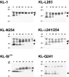
Biosynthetic characteristics of KL-1 cytoplasmic domain variants and KL-Sl 17H. COS-1 cells were transfected with various mutant constructs, KL-1, KL-L263, KL-N254, KL-Δ241/254, KL-Sl 17H, and KL-Q241, labeled with 35S-methionine for 30 min, and chased in regular medium. Cell lysates were collected at designated time points, immunoprecipitated with anti-KL antibody, and analyzed by SDS-PAGE (12%). Arrowheads, Mature glycosylated KL proteins. *Immature forms. Molecular mass (Mr) markers are indicated in kilodaltons.
Figure 3
Figure 3
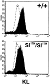
Determination of cell surface expression of KL in Sl 17H/Sl 17H fibroblasts by FACS®.
Figure 4
Figure 4
Endo H treatment of KL-1, KL cytoplasmic domain variants, and KL-Sl 17H. COS-1 cells were transfected with different KL constructs and labeled with 35S-methionine. Cell lysates collected at different time points were immunoprecipitated and treated in the presence or absence of endo H. The reaction products were analyzed by SDS-PAGE (12%). Arrowheads, Immature KL protein products.
Figure 4
Figure 4
Endo H treatment of KL-1, KL cytoplasmic domain variants, and KL-Sl 17H. COS-1 cells were transfected with different KL constructs and labeled with 35S-methionine. Cell lysates collected at different time points were immunoprecipitated and treated in the presence or absence of endo H. The reaction products were analyzed by SDS-PAGE (12%). Arrowheads, Immature KL protein products.
Figure 5
Figure 5
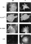
KL protein expression detected by immunofluorescence in COS-1 cells expressing wild-type and mutant KL protein products. COS-1 cells transfected with KL-1, KL-Q241, and KL-Sl 17H grown on coverslips were fixed with 2% formaldehyde (NP, nonpermeabilized control) and permeabilized with 0.1% saponin (P). The cells were incubated with anti-KL antibody or an ER marker antibody and FITC-conjugated goat anti– rabbit antibody. Photographs were taken with fluorescent microscopy.
Figure 6
Figure 6
Demonstration of KL-1, KL-2 dimers in transfected COS-1 cells. COS-1 cells were transfected with various normal and mutant KL-1 and KL-2 constructs: (A) KL-1, KL-2, and KL-S; (B) KL-1: L263, V248, Δ241/254; KL-2: L263, N254, Q241. 72 h after transfection, they were treated with the cross-linkers DSS and BS3 for 1 h at 37°C. Cell lysates were collected and fractionated by SDS-PAGE (12%), and KL protein products were identified by Western blotting. Arrowheads, KL dimers. *Monomers. Molecular mass (Mr) markers are indicated in kilodaltons.
Figure 7
Figure 7
The Sl 17H mutation affects dimer formation on the cell surface. COS-1 cells were transfected with normal KL-1 constructs and mutant Sl17H constructs containing various COOH-terminal deletions: Sl 17H-T257, Sl 17H-R249, and Sl 17H-A240. 72 h after transfection, cell surface proteins were biotinylated, and cells were treated with the cross-linkers DSS and BS3 (as indicated) for 1 h at 37°C. Cell lysates were fractionated on streptavidin-sepharose columns, the biotinylated fraction was electrophoretically fractionated by SDS-PAGE (12%,) and KL protein products were identified by Western blotting. Arrowheads, KL dimers. *Monomers. Molecular mass (Mr) markers are indicated in kilodaltons.
Figure 8
Figure 8
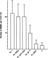
Adhesion of BMMCs to COS-1 cells expressing KL-1, secretory KL-1S, different KL cytoplasmic variants, and KL-Sl 17H. COS-1 cells were transferred to 24-well plates 24 h after transfection at a density of 7.5 × 10⁴ cells per well. BMMCs (1.5 × 10⁵ cells per well) were incubated with transfected COS-1 cells for 1 h in the presence and absence of anti–c-kit antibody. Nonattached BMMCs were washed away three times with medium, and the number of attached BMMCs was counted by microscopic inspection. The number of BMMCs attached to individual COS cells is shown.
Figure 9
Figure 9
Long-term culture of bone marrow cells from Sl 17H/Sl 17H mice. Nonadherent cells were counted at weekly intervals. Results from three separate experiments are shown.

Similar articles

Cited by

References

    1. Lee DC, Fenton SE, Berkowitz EA, Hissong MA. Transforming growth factor: expression, regulation, and biological activities. Pharmacol Rev. 1995;47:51–85. - PubMed
    1. Huang EJ, Nocka KH, Buck J, Besmer P. Differential expression and processing of two cell associated forms of the kit-ligand: KL-1 and KL-2. Mol Biol Cell. 1992;3:349–362. - PMC - PubMed
    1. Bosenberg MW, Massague J. Juxtacrine cell signaling molecules. Curr Opin Cell Biol. 1993;5:823–838. - PubMed
    1. Hill RJ, Sternberg PW. The gene lin-3 encodes an inductive signal for vulval development in C. elegans. . Nature. 1992;358:470–476. - PubMed
    1. Hart AC, Kramer H, Van Vacter DL, Jr, Paidhungat M, Zipursky SL. Induction of cell fate in the Drosophilaretina: the bride of sevenless protein is predicted to contain a large extracellular domain and seven transmembrane segments. Genes Dev. 1990;4:1835–1847. - PubMed

Publication types

MeSH terms

Substances