Comparative Study
. 1998 May;18(5):2462-73.
doi: 10.1128/MCB.18.5.2462.

The Gfi-1B proto-oncoprotein represses p21WAF1 and inhibits myeloid cell differentiation

Affiliations
Comparative Study

The Gfi-1B proto-oncoprotein represses p21WAF1 and inhibits myeloid cell differentiation

B Tong et al. Mol Cell Biol. 1998 May.

Abstract

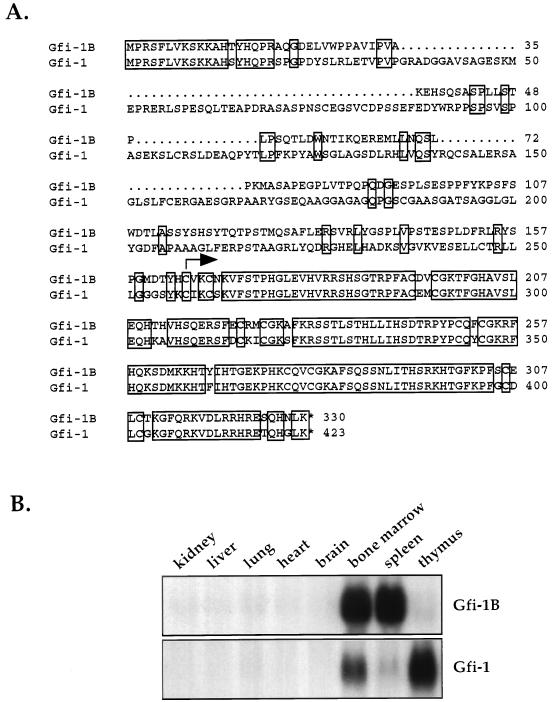
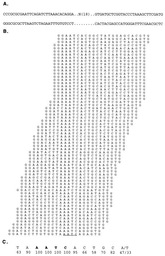
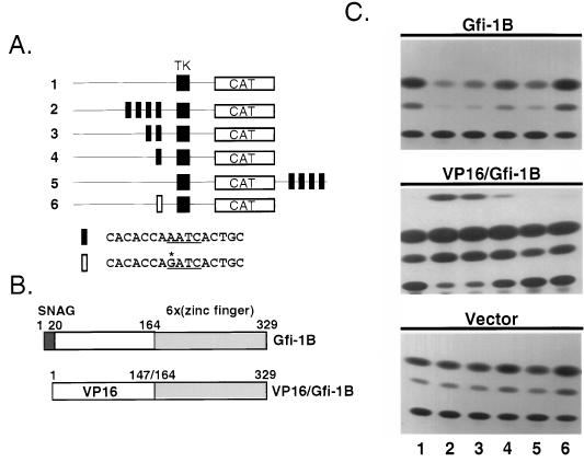
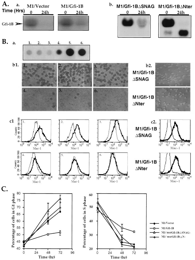
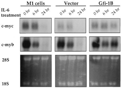
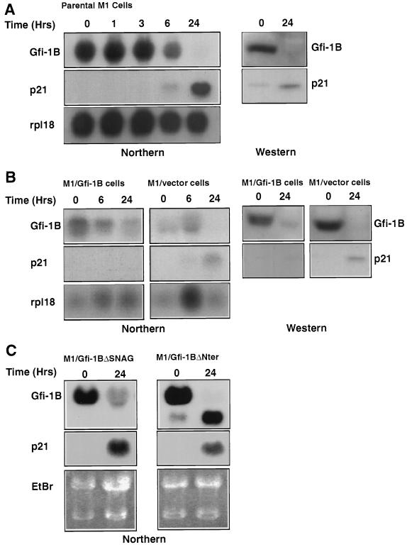
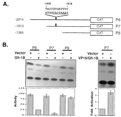
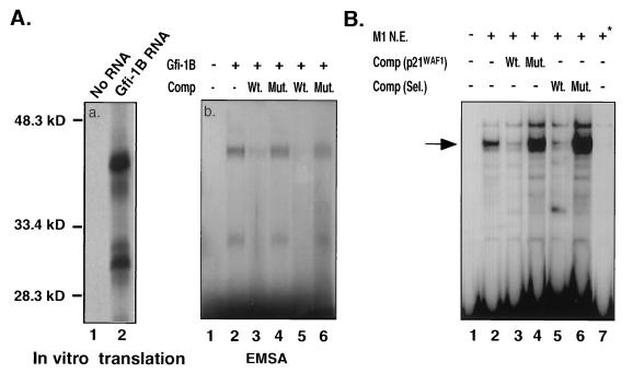
Gfi-1 is a cellular proto-oncogene that was identified as a target of provirus integration in T-cell lymphoma lines selected for interleukin-2 (IL-2) independence in culture and in primary retrovirus-induced lymphomas. Gfi-1 encodes a zinc finger protein that functions as a transcriptional repressor. Here we show that Gfi-1B, a Gfi-1 related gene expressed in bone marrow and spleen, also encodes a transcriptional repressor. IL-6-induced G1 arrest and differentiation of the myelomonocytic cell line M1 were linked to the downregulation of Gfi-1B and the parallel induction of the cyclin-dependent kinase inhibitor p21WAF1. Experiments addressing the potential mechanism of the apparent coordinate regulation of these genes revealed that Gfi-1B represses p21WAF1 directly by binding to a high-affinity site at -1518 to -1530 in the p21WAF1 promoter. Forced expression of Gfi-1B, but not of Gfi-1B deletion mutants lacking the repressor domain, blocked the IL-6-mediated induction of p21WAF1 and inhibited G1 arrest and differentiation. We conclude that Gfi-1B is a direct repressor of the p21WAF1 promoter, the first such repressor identified to date, and that sustained expression of Gfi-1B blocks IL-6-induced G1 arrest and differentiation of M1 cells perhaps because it prevents p21WAF1 induction by IL-6.


Figures

FIG. 1
FIG. 1
Gfi-1B encodes a Gfi-1-related zinc finger protein that is expressed in bone marrow and spleen. (A) Predicted amino acid sequence of Gfi-1B and comparison with Gfi-1. The two proteins exhibit a high degree of homology in the SNAG repressor domain (amino acids 1 to 19) and the zinc finger domain (amino acids 164 to 329). The arrow indicates the start of the zinc finger region. (B) Tissue distribution of Gfi-1B and Gfi-1. Gfi-1B is expressed in bone marrow and spleen, whereas Gfi-1 is expressed primarily in bone marrow and thymus.
FIG. 2
FIG. 2
Definition of the Gfi-1B binding site. (A) Double-stranded DNA oligonucleotides used for Gfi-1B binding and PCR amplification. The original oligonucleotide pool contained an 18-bp random sequence flanked by fixed sequences that were used for PCR amplification. (B) Alignment of 64 sequences of the selected oligonucleotides containing one AATC motif (underlined). (C) Deduced Gfi-1B binding site. The numbers show the frequencies of individual bases at each position.
FIG. 3
FIG. 3
Gfi-1B represses whereas a VP16/Gfi-1B chimera activates promoters containing a Gfi-1B binding site(s). (A) Schematic diagram of six different CAT reporter constructs. CAT expression by these constructs is under the control of a minimal herpes simplex virus thymidine kinase promoter (construct 1) with one (construct 4), two (construct 3) or four (construct 2) Gfi-1B binding sites placed upstream or four sites placed downstream (construct 5) of the CAT gene. Alternatively, one mutant site with a GATC instead of an AATC core motif was placed upstream of the thymidine kinase promoter (construct 6). (B) Schematic diagram of Gfi-1B and a VP16/Gfi-1B chimera. In this chimera the Gfi-1B amino-terminal region (amino acids 1 to 164) was replaced by the acidic activation domain of VP16 (amino acids 1 to 147). (C) Cotransfection of the six CAT reporter constructs (1.8 μg) shown in panel A with a CMV5/Gfi-1B construct (100 ng) into NIH 3T3 cells results in repression of CAT gene expression (top panel, lanes 2 to 5; the lane number corresponds to the number of the reporter construct used in that lane, as shown in panel A), while cotransfection of the same reporter constructs with the CMV5 empty vector (100 ng) does not (bottom panel). Cotransfection of the reporter constructs with a CMV5 expression construct of the VP16/Gfi-1B chimera (100 ng) shown in panel B induces CAT expression from the same constructs that were repressed by Gfi-1B (middle panel). Mutation of the AATC motif to GATC abrogates both the repression and the induction of CAT expression (lane 6).
FIG. 4
FIG. 4
Forced expression of Gfi-1B inhibits IL-6-induced G1 arrest and differentiation of the murine myeloblastic leukemia cell line M1. (Aa) Northern blot of total-cell RNA derived from clones of M1 cells transfected with the pcDNA3 vector (M1/vector) or a pcDNA3 expression construct of Gfi-1B (M1/Gfi-1B) probed with a Gfi-1B cDNA probe. Cells were harvested before (time 0) and 24 h after treatment with IL-6. (Ab) Northern blot of total-cell RNA derived from M1 cells stably transfected with pcDNA3-based expression constructs of Gfi-1BΔSNAG or Gfi-1BΔNter probed with a Gfi-1B cDNA probe. Cells were also harvested before (time 0) and 24 h after treatment with IL-6. (Ba) Gfi-1B expression in six M1 cell clones 24 h after exposure to IL-6. Clone 1 was untransfected parental M1, clone 2 was transfected with pcDNA3, and clones 3 to 6 were transfected with the pcDNA3/Gfi-1B construct. (Bb1 and Bc1) Constitutive expression of Gfi-1B blocks IL-6-induced differentiation of M1 cells. Differentiation was assessed by measuring the relative cell attachment to the surface of the petri dish at 48 h after IL-6 treatment (b1) and Mac-1 expression at 96 h after IL-6 treatment (c1). The superimposed graphs in panel Bc depict fluorescent intensity versus cell number in untreated and IL-6-treated cell cultures. Graphs 1 to 6 in both panels Bb1 and Bc1 correspond to the six M1 clones analyzed for Gfi-1B expression after the IL-6 treatment shown in panel Ba. (Bb2 and Bc2) M1 cells stably transfected with Gfi-1BΔSNAG or Gfi-1BΔNter do not differentiate in response to IL-6 as determined by cell attachment to plastic (Bb2) and by Mac1 expression (Bc2). Identical results were obtained from four independent cultures of M1 cells expressing these mutants. (C) Constitutive expression of Gfi-1B in M1 cells inhibits IL-6-induced G1 arrest. M1/vector, M1/Gfi-1B, M1/mutGfi-1B(ΔSNAG), and M1/mutGfi-1B(ΔN) cells were examined for cell cycle distribution at 0, 48, and 72 h after IL-6 treatment. Each percentage of cells in G1 or S represents an average of three independent experiments.
FIG. 5
FIG. 5
Epistatic relationship of Gfi-1B, c-myc, and c-myb during IL-6-induced differentiation of M1 cells. Northern blots of total RNA from parental M1 cells and pcDNA3 and pcDNA3/Gfi-1B-transfected M1 cells were hybridized to c-myc and c-myb probes. Cells were harvested at 0, 6, and 24 h following exposure to IL-6. Ethidium bromide staining of the RNA is presented as a measure of loading.
FIG. 6
FIG. 6
Induction of p21WAF1 during IL-6-induced differentiation of M1 cells depends on the downregulation of Gfi-1B. (A, left) Northern blots of total-cell RNA from parental M1 cells harvested at the indicated time points following IL-6 treatment. The blot was hybridized to a probe derived from the unique non-zinc finger region of Gfi-1B (upper panels), a p21WAF1 probe derived from the coding region of the gene (middle panel), and the ribosomal protein rpl18 (lower panel). (A, right) Western blots of lysates from the same cells, harvested at 0 and 24 h after treatment with IL-6 and probed with the anti-Gfi-1B antiserum (upper panel) or with the anti-p21 antibody (lower panel). (B, left) Northern blots of M1 cells stably transfected with pcDNA3 or pcDNA3/Gfi-1B and harvested at the indicated time points following treatment with IL-6. The blots were hybridized to the same Gfi-1B probe (upper panels), p21WAF1 probe (middle panels), or ribosomal protein probe (lower panels) listed for panel A. (B, right) Western blots of lysates from the cells shown on the left side of panel B probed with anti-Gfi-1B (upper panel) or anti-p21WAF1 (lower panel) antibodies. (C) Forced expression of Gfi-1BΔSNAG or Gfi-1BΔNter constructs in M1 cells fails to inhibit IL-6-mediated induction of p21WAF1. Northern blots of M1 cells stably transfected with pcDNA3-based expression constructs of Gfi-1BΔSNAG or Gfi-1BΔNter and harvested at the indicated time points following treatment with IL-6 are shown. The blots were probed with Gfi-1B (upper panels) or p21WAF1 (middle panels) probes. RNA loading was determined by staining the gel with ethidium bromide (lower panel).
FIG. 7
FIG. 7
Gfi-1B is a direct repressor of the p21WAF1 promoter. (A) Schematic diagram of sequentially deleted p21WAF1 promoter-CAT reporter constructs. The location and sequence of a putative Gfi-1B binding site at −1530 to −1518 are marked. (B) Gfi-1B represses whereas the VP16/Gfi-1B chimera activates the p21WAF1 promoter. The p53-deficient mouse embryonic fibroblast cell line 10-1 was cotransfected as indicated with the reporter constructs shown in panel A and Gfi-1B (left panels) or the VP16/Gfi-1B chimera (right panels). Repression and activation were quantitatively measured with a phosphorimager. Repression measurements were based on three independent experiments. Activation measurements were based on two independent experiments.
FIG. 8
FIG. 8
Gfi-1B binds the p21WAF1 promoter directly. (Aa) [35S]methionine-labeled, in vitro-translated Gfi-1B was electrophoresed in 10% SDS-PAGE. Lanes: 1, products of in vitro translation carried out in the absence of exogenously added RNA; 2, in vitro translation in the presence of Gfi-1B RNA. The lower band is likely to represent a Gfi-1B product translated from an internal AUG. (Ab) A 28-bp double-stranded oligonucleotide containing the putative Gfi-1B binding site of the p21WAF1 promoter was 32P labeled and used as a probe in an EMSA. Lane 1 contains the oligonucleotide probe and reticulocyte lysate only (negative control). Comp, oligonucleotide competition; Wt and Mut, wild-type and mutant oligonucleotide competitors, respectively. In lanes 3 and 4, the oligonucleotide competitors were added at a 100-fold excess, whereas in lanes 5 and 6 they were added at a 200-fold excess. (B) The same double-stranded oligonucleotide probe used in panel A was incubated with nuclear extracts from untreated (lanes 2 to 6) or IL-6-treated (lane 7) M1 cells. The major shifted band, indicated by an arrow, was competed by excess wild-type oligonucleotide competitors comp (p21WAF1) derived from the p21WAF1 promoter, and the Comp (Sel.) band was derived via the oligonucleotide selection experiment (lanes 3 and 5). Nonbinding mutants of the same oligonucleotides did not compete (lanes 4 and 6).

References

    1. Anderson S, Davis D H, Dahlbäck H, Jörnvall H, Russell D W. Cloning, structure and expression of the mitochondrial cytochrome P-450 sterol 26-hydroxylase, a bile biosynthetic enzyme. J Biol Chem. 1989;264:8222–8229. - PubMed
    1. Arnaout M A. Structure and function of the leukocyte adhesion molecules CD11/CD18. Blood. 1990;75:1037–1050. - PubMed
    1. Bell D W, Taguchi T, Jenkins N A, Gilbert D J, Copeland N G, Gilks C B, Zweidler-McKay P, Grimes H L, Tsichlis P N, Testa J R. Chromosomal localization of a gene, GFI1, encoding a novel zinc finger protein reveals a new syntenic region between man and rodents. Cytogenet Cell Genet. 1995;70:263–267. - PubMed
    1. Bies J, Hoffman B, Amanullah A, Giese T, Wolff L. B-Myb prevents growth arrest associated with terminal differentiation of monocytic cells. Oncogene. 1996;12:355–363. - PubMed
    1. Biggs J R, Kudlow J E, Kraft A S. The role of the transcription factor Sp1 in regulating the expression of the WAF1/CIP1 gene in U937 leukemic cells. J Biol Chem. 1996;271:901–906. - PubMed

Publication types

MeSH terms

Associated data