. 1998 May;18(5):2474-85.
doi: 10.1128/MCB.18.5.2474.

Wnt-1 induces growth, cytosolic beta-catenin, and Tcf/Lef transcriptional activation in Rat-1 fibroblasts

Affiliations

Wnt-1 induces growth, cytosolic beta-catenin, and Tcf/Lef transcriptional activation in Rat-1 fibroblasts

C S Young et al. Mol Cell Biol. 1998 May.

Abstract

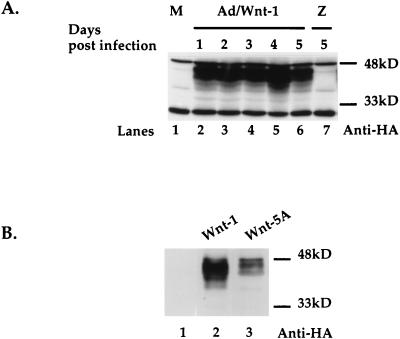
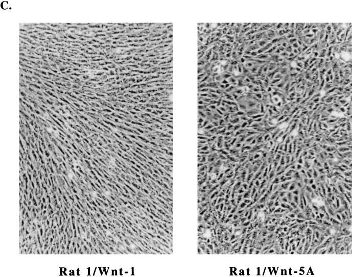
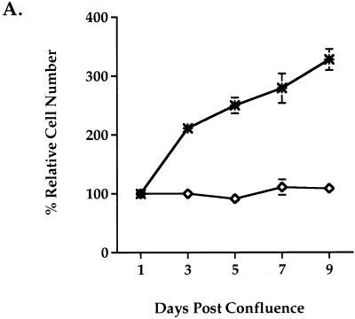
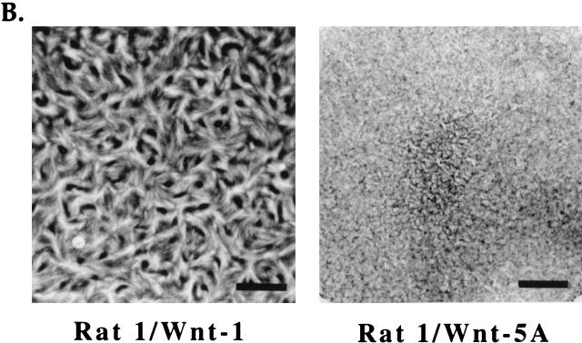
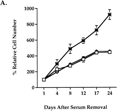
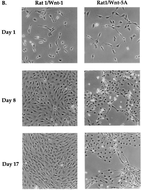
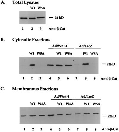
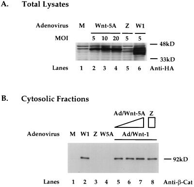
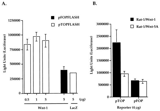
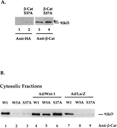
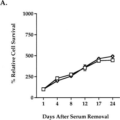
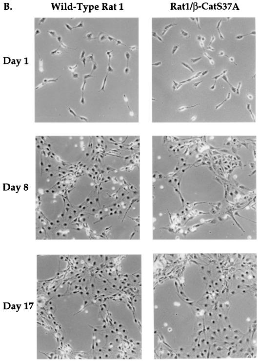
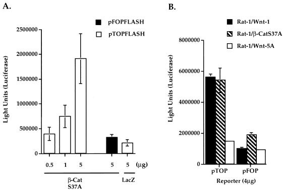
Genetic evidence suggests that regulation of beta-catenin and regulation of Tcf/Lef family transcription factors are downstream events of the Wnt signal transduction pathway. However, a direct link between Wnt activity and Tcf/Lef transcriptional activation has yet to be established. In this study, we show that Wnt-1 induces a growth response in a cultured mammalian cell line, Rat-1 fibroblasts. Wnt-1 induces serum-independent cellular proliferation of Rat-1 fibroblasts and changes in morphology. Rat-1 cells stably expressing Wnt-1 (Rat-1/Wnt-1) show a constitutive up-regulation of cytosolic beta-catenin, while membrane-associated beta-catenin remains unaffected. Induction of cytosolic beta-catenin in Rat-1/Wnt-1 cells is correlated with activation of a Tcf-responsive transcriptional element. We thus provide evidence that Wnt-1 induces Tcf/Lef transcriptional activation in a mammalian system. Expression of a mutant beta-catenin (beta-CatS37A) in Rat-1 cells does not result in a proliferative response or a detectable change in the cytosolic beta-catenin protein level. However, beta-CatS37A expression in Rat-1 cells results in strong Tcf/Lef transcriptional activation, comparable to that seen in Wnt-1-expressing cells. These results suggest that Wnt-1 induction of cytosolic beta-catenin may have functions in addition to Tcf/Lef transcriptional activation.


Figures

FIG. 1
FIG. 1
Wnt-1 expression alters Rat-1 fibroblast morphology and growth. (A) Transient expression of Wnt-1 protein by recombinant adenovirus expression vector in Rat-1 fibroblasts. Western blot analysis of cell lysates shows Wnt-1 protein expressed in cells at multiple days (days 1 to 5) after infection with Ad/Wnt-1 (lanes 2 to 6) (MOI = 5) but not with the control, Ad/LacZ (Z) (MOI = 5) (lane 7), or after mock infection (M) (lane 1). Wnt-1 was detected with anti-HA antibody (Anti-HA). (B) Stable expression of Wnt-1 and Wnt-5A proteins in Rat-1 stable cell lines, Rat-1/Wnt-1 and Rat-1/Wnt-5A. Western blot analysis of cell lysates shows Wnt-1 (lane 2) and Wnt-5A (lane 3) proteins expressed, as compared to control cells (lane 1). Wnt-1 and Wnt-5A proteins were detected with anti-HA antibody. (C) Wnt-1 alters Rat-1 fibroblast morphology and growth. Phase-contrast microscopy of polyclonal Rat-1/Wnt-1 and Rat-1/Wnt-5A stable cell lines shows that Rat-1/Wnt-1 cells attain an elongated, refractile morphology and densely pack in cord-like bundles, in contrast to Rat-1/Wnt-5A cells.
FIG. 1
FIG. 1
Wnt-1 expression alters Rat-1 fibroblast morphology and growth. (A) Transient expression of Wnt-1 protein by recombinant adenovirus expression vector in Rat-1 fibroblasts. Western blot analysis of cell lysates shows Wnt-1 protein expressed in cells at multiple days (days 1 to 5) after infection with Ad/Wnt-1 (lanes 2 to 6) (MOI = 5) but not with the control, Ad/LacZ (Z) (MOI = 5) (lane 7), or after mock infection (M) (lane 1). Wnt-1 was detected with anti-HA antibody (Anti-HA). (B) Stable expression of Wnt-1 and Wnt-5A proteins in Rat-1 stable cell lines, Rat-1/Wnt-1 and Rat-1/Wnt-5A. Western blot analysis of cell lysates shows Wnt-1 (lane 2) and Wnt-5A (lane 3) proteins expressed, as compared to control cells (lane 1). Wnt-1 and Wnt-5A proteins were detected with anti-HA antibody. (C) Wnt-1 alters Rat-1 fibroblast morphology and growth. Phase-contrast microscopy of polyclonal Rat-1/Wnt-1 and Rat-1/Wnt-5A stable cell lines shows that Rat-1/Wnt-1 cells attain an elongated, refractile morphology and densely pack in cord-like bundles, in contrast to Rat-1/Wnt-5A cells.
FIG. 2
FIG. 2
Wnt-1 induces proliferation in quiescent Rat-1 fibroblasts. Rat-1/Wnt-1 (∗) and control Rat-1/Wnt-5A (◊) cells were cultured to confluence, deprived of serum, and monitored for growth postconfluence. (A) At multiple days postconfluence, viable cells were trypsinized and counted by the trypan blue exclusion method. The percent relative cell number is calculated as the percentage relative to the cell number recorded 1 day after serum removal. Triplicate samples were counted and averaged. (B) Six days postconfluence, cell monolayers were fixed and stained with Giemsa nuclear stain and photographed. Bar, 4.75 mm.
FIG. 2
FIG. 2
Wnt-1 induces proliferation in quiescent Rat-1 fibroblasts. Rat-1/Wnt-1 (∗) and control Rat-1/Wnt-5A (◊) cells were cultured to confluence, deprived of serum, and monitored for growth postconfluence. (A) At multiple days postconfluence, viable cells were trypsinized and counted by the trypan blue exclusion method. The percent relative cell number is calculated as the percentage relative to the cell number recorded 1 day after serum removal. Triplicate samples were counted and averaged. (B) Six days postconfluence, cell monolayers were fixed and stained with Giemsa nuclear stain and photographed. Bar, 4.75 mm.
FIG. 3
FIG. 3
Wnt-1 induces serum-independent growth in Rat-1 fibroblasts. Wild-type Rat-1 (□), Rat-1/Wnt-1 (∗), and control Rat-1/Wnt-5A (◊) cells were seeded at low density (<15% confluence), deprived of serum, and monitored for serum-independent growth. (A) At multiple days after removal of serum, the cells were trypsinized and viable cells were counted. The percent relative cell number is calculated relative to the cell number recorded 1 day after serum removal. Triplicate samples were counted and averaged. (B) At multiple days (days 1, 8, and 17) after removal of serum, Rat-1/Wnt-1 and Rat-1/Wnt-5A cells were photographed and monitored for serum-independent growth.
FIG. 3
FIG. 3
Wnt-1 induces serum-independent growth in Rat-1 fibroblasts. Wild-type Rat-1 (□), Rat-1/Wnt-1 (∗), and control Rat-1/Wnt-5A (◊) cells were seeded at low density (<15% confluence), deprived of serum, and monitored for serum-independent growth. (A) At multiple days after removal of serum, the cells were trypsinized and viable cells were counted. The percent relative cell number is calculated relative to the cell number recorded 1 day after serum removal. Triplicate samples were counted and averaged. (B) At multiple days (days 1, 8, and 17) after removal of serum, Rat-1/Wnt-1 and Rat-1/Wnt-5A cells were photographed and monitored for serum-independent growth.
FIG. 4
FIG. 4
Wnt-1 induces cytosolic accumulation of β-catenin protein in Rat-1 fibroblasts. Western blot analyses of steady-state β-catenin protein levels in unfractionated (A), cytosolic (B), and membranous (C) protein fractions of wild-type Rat-1 (blank), Rat-1/Wnt-1 (W1), and Rat-1/Wnt-5A (W5A) cells are shown. Cells were uninfected or infected with Ad/Wnt-1 or with the control, Ad/LacZ (MOI = 5). Cell extracts were prepared and fractionated, as described in Materials and Methods. Protein fractions were separated by SDS-PAGE (10% polyacrylamide), transferred onto nitrocellulose, and analyzed by Western blot analysis with anti-β-catenin antibody (Anti-β-Cat).
FIG. 5
FIG. 5
Coexpression of Wnt-5A does not interfere Wnt-1 induction of cytosolic β-catenin in Rat-1 fibroblasts. (A) Expression of Wnt-5A protein in Rat-1 cells at increasing MOIs with Ad/Wnt-5A. Western blot analysis of cell lysates 2 days after infection with Ad/Wnt-5A at an MOI of 5, 10, or 20 or with Ad/Wnt-1 (W1) (MOI = 5) or Ad/LacZ (Z) (MOI = 5) or after mock infection (M) is shown. Wnt-1 and Wnt-5A proteins are detected with anti-HA antibody. (B) Coexpression of Wnt-5A does not interfere with Wnt-1 induction of cytosolic β-catenin in Rat-1 fibroblasts. Western blot analyses of steady-state cytosolic β-catenin protein levels in Rat-1 cells following mock infection (M), infection with Ad/Wnt-1 (W1), Ad/LacZ (Z), or Ad/Wnt-5A (5A), coinfection with Ad/Wnt-1 (MOI = 5) and Ad/Wnt-5A (MOI = 5, 10, or 20), or coinfection with Ad/Wnt-1 (W1) (MOI = 5) and Ad/LacZ (Z) (MOI = 20) are shown. Cell extracts were prepared, fractionated, and analyzed by Western blot analysis with anti-β-catenin antibody (Anti-β-Cat).
FIG. 6
FIG. 6
Wnt-1 induces Tcf/Lef-dependent transcription in Rat-1 fibroblasts. (A) Transient expression of Wnt-1 induces Tcf/Lef-dependent transcription. Wild-type (pTOPFLASH) or mutant (pFOPFLASH) Tcf reporter constructs (4 μg) were cotransfected into Rat-1 fibroblasts with increasing amounts of Wnt-1 or lacZ cDNA. (B) Stable expression of Wnt-1 in Rat-1/Wnt-1 cells induces constitutive Tcf/Lef transcriptional activation. Wild-type Tcf (pTOP) or mutant Tcf (pFOP) reporter constructs (4 μg) were transfected into Rat-1/Wnt-1 or Rat-1/Wnt-5A stable cell lines. Luciferase activities were measured 2 days after transfection. Triplicate samples were counted and averaged.
FIG. 7
FIG. 7
Mutant β-catenin (S37A) does not accumulate in the cytosol. (A) Western blot analyses of β-catenin (S37A) in Rat-1/β-CatS37A stable cell lines. Cell lysates were separated by SDS-PAGE (10% polyacrylamide for lanes 1 and 2 and 7.5% polyacrylamide for lanes 3 and 4) and transferred to nitrocellulose, and β-catenin (S37A) was detected by Western blot analysis with anti-HA antibody (lane 2) or anti-β-catenin (lane 4). Lane 1 shows that no exogenous protein is detected in control cells. Lane 3 shows endogenous expression of β-catenin in Rat-1 cells. Exogenous β-catenin (S37A) protein is indicated by the arrow in lane 4. (B) Western blot analysis of cytosolic β-catenin protein levels in Rat-1/Wnt-1 (W1), Rat-1/Wnt-5A (W5A), and Rat-1/β-CatS37A (S37A) stable cell lines. Cells were uninfected or infected with Ad/Wnt-1 (MOI = 5) or with the control, Ad/LacZ (MOI = 5) Cytosolic protein fractions were separated by SDS-PAGE (10% polyacrylamide), transferred to nitrocellulose, and analyzed by Western blot analysis with anti-β-catenin antibody (Anti-β-Cat).
FIG. 8
FIG. 8
Mutant β-catenin (S37A) expression does not alter Rat-1 cell morphology or growth properties. Rat-1/β-CatS37A (◊) and wild-type Rat-1 (□) cells were seeded at low density (<15% confluence), deprived of serum, and monitored for serum-independent growth. (A) At multiple days after removal of serum, the cells were trypsinized and viable cells were counted. The percent relative cell number is calculated as the percentage relative to the cell number recorded 1 day after serum removal. Samples were counted in triplicate. (B) After removal of serum, wild-type Rat-1 and Rat-1/β-CatS37A cells were photographed on the days indicated in the figure.
FIG. 8
FIG. 8
Mutant β-catenin (S37A) expression does not alter Rat-1 cell morphology or growth properties. Rat-1/β-CatS37A (◊) and wild-type Rat-1 (□) cells were seeded at low density (<15% confluence), deprived of serum, and monitored for serum-independent growth. (A) At multiple days after removal of serum, the cells were trypsinized and viable cells were counted. The percent relative cell number is calculated as the percentage relative to the cell number recorded 1 day after serum removal. Samples were counted in triplicate. (B) After removal of serum, wild-type Rat-1 and Rat-1/β-CatS37A cells were photographed on the days indicated in the figure.
FIG. 9
FIG. 9
Mutant β-catenin (S37A) activates Tcf/Lef-dependent transcription. (A) Transient expression of β-catenin (S37A) (β-CatS37A) induces Tcf/Lef-dependent transcription. Wild-type (pTOPFLASH) or mutant (pFOPFLASH) Tcf reporter constructs (4 μg) were cotransfected into Rat-1 fibroblasts with increasing amounts of β-CatS37A or control lacZ cDNAs. (B) Stable expression of β-CatS37A in Rat-1/β-CatS37A cells induces constitutive Tcf/Lef transcriptional activation. Wild-type Tcf (pTOP) or mutant Tcf (pFOP) reporter constructs (4 μg) were transfected into Rat-1/β-CatS37A, Rat-1/Wnt-1, or Rat-1/Wnt-5A stable cell lines. Luciferase activities were measured 2 days after transfection. Triplicate samples were counted and averaged.

References

    1. Aberle H, Bauer A, Stappert J, Kispert A, Kemler R. β-Catenin is a target for the ubiquitin-proteasome pathway. EMBO J. 1997;16:3797–3804. - PMC - PubMed
    1. Behrens J, von Kries J P, Kuhl M, Bruhn L, Wedlich D, Grosschedl R, Birchmeier W. Functional interaction of beta-catenin with the transcription factor LEF-1. Nature. 1996;382:638–642. - PubMed
    1. Bett A J, Haddara W, Prevec L, Graham F L. An efficient and flexible system for construction of adenovirus vectors with insertions or deletions in early regions 1 and 3. Proc Natl Acad Sci USA. 1994;91:8802–8806. - PMC - PubMed
    1. Bhanot P, Brink M, Samos C H, Hsieh J-C, Wang Y, Macke J P, Andrew D, Nathans J, Nusse R. A new member of the frizzled family from Drosophila functions as a Wingless receptor. Nature. 1996;382:225–230. - PubMed
    1. Bradbury J M, Niemeyer C C, Dale T C, Edwards P A W. Alterations of the growth characteristics of the fibroblast cell line C3H10T1/2 by members of the Wnt gene family. Oncogene. 1994;9:2597–2603. - PubMed

Publication types

LinkOut - more resources