. 1998 May;18(5):2545-52.
doi: 10.1128/MCB.18.5.2545.

Stat3 activation by Src induces specific gene regulation and is required for cell transformation

Affiliations

Stat3 activation by Src induces specific gene regulation and is required for cell transformation

J Turkson et al. Mol Cell Biol. 1998 May.

Abstract

While signal transducers and activators of transcription (STATs) were originally discovered as intracellular effectors of normal signaling by cytokines, increasing evidence also points to a role for STAT transcription factors in oncogenesis. Previous studies have demonstrated that one STAT family member, Stat3, possesses constitutively elevated tyrosine phosphorylation and DNA-binding activity in fibroblasts stably transformed by the Src oncoprotein. To determine if this Stat3 activation by Src could induce Stat3-mediated gene expression, luciferase reporter constructs based on synthetic and authentic promoters were transfected into NIH 3T3 cells. Activation of endogenous cellular Stat3 by the Src oncoprotein induced gene expression through a Stat3-specific binding element (TTCCCGAA) of the C-reactive protein gene promoter. A naturally occurring splice variant of human Stat3 protein, Stat3beta, with a deletion in the C-terminal transactivation domain abolished this gene induction in a dominant negative manner. Expression of Stat3beta did not have any effect on a reporter construct based on the c-fos serum response element, which is not dependent on Stat3 signaling, indicating that Stat3beta does not nonspecifically inhibit other signaling pathways or Src function. Transfection of vectors expressing Stat3beta together with Src blocked cell transformation by Src as measured in a quantitative focus formation assay using NIH 3T3 cells. By contrast, Stat3beta had a much less pronounced effect on focus formation induced by the Ras oncoprotein, which does not activate Stat3 signaling. In addition, three independent clones of NIH 3T3 cells stably overexpressing Stat3beta were generated and characterized, demonstrating that Stat3beta overexpression does not have a toxic effect on cell viability. These Stat3beta-overexpressing clones were shown to be deficient in Stat3-mediated signaling and refractory to Src-induced cell transformation. We conclude that Stat3 activation by the Src oncoprotein leads to specific gene regulation and that Stat3 is one of the critical signaling pathways involved in Src oncogenesis. Our findings provide evidence that oncogenesis-associated activation of Stat3 signaling is part of the process of malignant transformation.


Figures
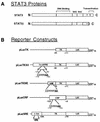
FIG. 1
FIG. 1
Schematic representations of Stat3 proteins and reporter constructs. (A) Full-length Stat3 and Stat3β, which is a naturally occurring splice variant with a deletion in the C-terminal transactivation domain, are diagrammed with various protein domains and the major sites of phosphorylation shown. (B) The reporter construct pLucTK contains only the TK minimal promoter driving luciferase (LUC) gene expression, while pLucTKS3 has seven copies of a Stat3-specific binding site (cAPRE) from the CRP gene inserted upstream of the TK minimal promoter. pLucCRP contains an authentic CRP promoter fragment driving expression of the luciferase gene. pLucTKSIE has two copies of a high-affinity mutant (hSIE) of the c-fos SIE inserted upstream of the TK minimal promoter, whereas pLucSRE contains a c-fos promoter fragment harboring the SRE inserted upstream of TK promoter sequences. Not shown is the basic pLuc backbone vector, which contains the luciferase gene without promoter sequences cloned in pUC19. See Materials and Methods for additional details of constructs.
FIG. 2
FIG. 2
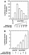
Induction of Stat3-specific gene expression by v-Src. NIH 3T3 cells were transiently transfected with the indicated plasmid vectors, and luciferase reporter activity in cytosolic extracts was measured as light emission, with a luminometer. (A) Cells were transfected with pLucTK reporter alone or pLucTKS3 reporter plus increasing concentrations of the v-Src expression vector, pMvsrc. (B) Cells were transfected with pLucTK or pLucTKS3 reporters in the presence or absence of vectors encoding v-Src, Stat3β, or both. Values shown in each panel are means plus standard deviations of at least four independent transfections, each performed in triplicate. For each transfection, luciferase activity was normalized to transfection efficiency, with β-Gal activity as an internal control.
FIG. 3
FIG. 3
Stat3β disrupts, and Stat3 augments, Src-induced gene expression. NIH 3T3 cells were transiently transfected and luciferase reporter activities were assayed as described for Fig. 1. (A) Cells were transfected with the Stat3 reporter, pLucTKS3, and v-Src vector together with increasing concentrations of vector encoding Stat3β. (B) Cells were transfected with pLucTKS3 reporter and vectors encoding v-Src, full-length Stat3 (pVRStat3), or both. Values are means plus standard deviations of at least three independent experiments, each performed in triplicate and normalized to β-Gal activity.
FIG. 4
FIG. 4
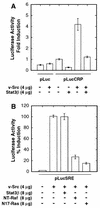
Stat3β specifically blocks Stat3 but not Ras or Raf-1 signaling induced by v-Src. NIH 3T3 cells were transiently transfected and luciferase reporter activities were assayed as described for Fig. 1. (A) Cells were transfected with pLucCRP reporter together with or without vectors encoding v-Src, Stat3β, or both. (B) Cells were transfected with pLucSRE reporter together with v-Src in the presence or absence of vectors encoding Stat3β, N17-Ras, or NT-Raf. The N17-Ras and NT-Raf proteins are dominant negative mutants of c-H-Ras and Raf-1, respectively. Values are means plus standard deviations of at least three independent transfections, each performed in triplicate and normalized to β-Gal activity.
FIG. 5
FIG. 5
Specificity of promoter elements for STAT signaling induced by v-Src or IFN-γ. NIH 3T3 cells were transiently transfected and luciferase reporter activities were assayed as described for Fig. 1. (A) Cells were transfected with pLucTKSIE together with expression vectors for v-Src, Stat3β, or both. Transfectants with reporter alone or reporter and Stat3β vector were treated with IFN-γ for 5 h prior to harvest of cells. (B) Cells were transfected with the indicated reporters in the presence or absence of v-Src expression vector. Cells transfected with reporter alone were treated with IFN-γ for 5 h prior to harvest. Values are means plus standard deviations of at least three independent experiments, each performed in triplicate and normalized to β-Gal activity.
FIG. 6
FIG. 6
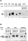
Induction of SIE binding activity by v-Src in transfected cells. Nuclear extracts were prepared from transiently transfected NIH 3T3 cells, and volumes containing equal amounts of total protein were subjected to EMSA by using 32P-labeled hSIE. (A) Cells were transfected with v-Src vector alone (NIH 3T3) or v-Src vector together with either Stat3 or Stat3β vector, as indicated. Lanes 7 to 10 are 1:10 dilutions of the samples loaded in lanes 3 to 6, respectively. Competitions of endogenous hSIE binding activity present in nuclear extracts of NIH 3T3 cells transfected with v-Src vector alone (lanes 12 and 13) were performed with a 100-fold molar excess of unlabeled hSIE or the unrelated c-fos intragenic regulatory element (FIRE) oligonucleotides. Supershifts (lanes 14 and 15) were performed with antibodies recognizing either amino acids 688 to 710 of Stat1 (αST1) or amino acids 750 to 769 of full-length Stat3 (αST3). (B) Nuclear extracts from cells transfected with v-Src vector plus Stat3 and/or Stat3β vector were subjected to EMSA, with competitions and supershifts performed as described for panel A. ST3, ST3β, and ST3/ST3β indicate migration of complexes containing Stat3 homodimers, Stat3β homodimers, and Stat3-Stat3β heterodimers, respectively. Asterisks indicate positions of supershifted complexes.
FIG. 7
FIG. 7
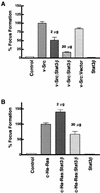
Cotransfection of Stat3β vector blocks transformation of NIH 3T3 cells induced by Src. (A) Cells were transfected with carrier DNA alone (control), with v-Src vector (200 ng) in the presence or absence of Stat3β vector (2 μg or 20 μg, as indicated) or empty vector, or with Stat3β vector (20 μg) alone. (B) Cells were transfected as described for panel A, except that activated c-H-Ras vector was used instead of v-Src vector. At least three independent sets of transfections were analyzed for Src and Ras focus formation assays. Values are means plus standard deviations of transfections from each experiment; percent focus formation is relative to that induced by Src or Ras alone (100%) within each of the independent sets of experiments.
FIG. 8
FIG. 8
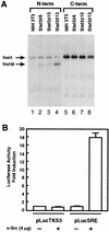
Characterization of NIH 3T3 cell lines stably overexpressing Stat3β. (A) Western blot analysis of whole-cell lysates prepared from three independent NIH 3T3 cell lines stably overexpressing Stat3β. Lanes 1 to 4 were probed with antibodies against the N-terminal portion of Stat3 which recognize both full-length Stat3 and Stat3β. Lanes 5 to 8 were probed with antibodies to the Stat3 C terminus which recognize full-length Stat3 but not Stat3β. (B) Clone Stat3β10 was transiently transfected with either pLucSRE or pLucTKS3 reporter in the presence or absence of vector encoding v-Src, as indicated. Values for luciferase activity are means plus standard deviations of at least two independent transfections, each performed in triplicate. For each transfection, luciferase activity was normalized to transfection efficiency by using β-Gal activity as an internal control.
FIG. 9
FIG. 9
Cell lines stably overexpressing Stat3β are resistant to cell transformation by Src. Focus formation assays were performed with normal NIH 3T3 cells or the three independent clones overexpressing Stat3β represented in Fig. 8. In each experiment, cells were transfected with 200 ng of v-Src expression vector, with the exception of Stat3β13, in which 20 μg of v-Src vector was used. Values are means plus standard deviations of three independent transfections, except in the case of Stat3β13, which was tested once.

References

    1. Aftab D T, Kwan J, Martin G S. Ras-independent transformation by v-Src. Proc Natl Acad Sci USA. 1997;94:3028–3033. - PMC - PubMed
    1. Akira S, Nishio Y, Inoue M, Wang X-J, Wei S, Matsusaka T, Yoshida K, Sudo T, Naruto M, Kishimoto T. Molecular cloning of APRF, a novel IFN-stimulated gene factor 3 p91-related transcription factor involved in the gp130-mediated signaling pathway. Cell. 1994;77:63–71. - PubMed
    1. Bowman, T., and R. Jove. Unpublished data.
    1. Bromberg J F, Horvath C M, Besser D, Lathem W W, Darnell J E., Jr Stat3 activation is required for cellular transformation by v-src. Mol Cell Biol. 1998;18:2553–2558. - PMC - PubMed
    1. Bromberg J F, Horvath C M, Wen Z, Schreiber R D, Darnell J E., Jr Transcriptionally active Stat1 is required for the antiproliferative effects of both interferon alpha and interferon gamma. Proc Natl Acad Sci USA. 1996;93:7673–7678. - PMC - PubMed

Publication types