. 1998 May;18(5):2608-16.
doi: 10.1128/MCB.18.5.2608.

The second catalytic domain of protein tyrosine phosphatase delta (PTP delta) binds to and inhibits the first catalytic domain of PTP sigma

Affiliations

The second catalytic domain of protein tyrosine phosphatase delta (PTP delta) binds to and inhibits the first catalytic domain of PTP sigma

M J Wallace et al. Mol Cell Biol. 1998 May.

Abstract

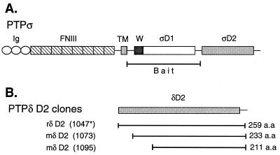
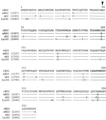
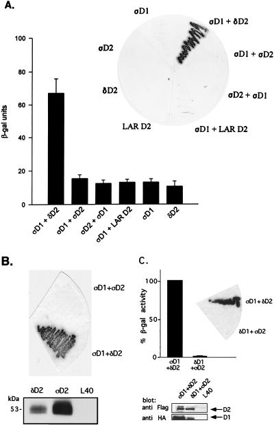
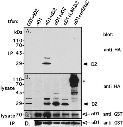
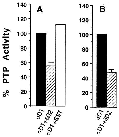
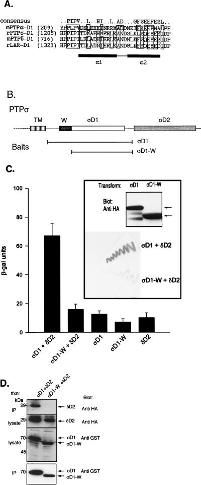
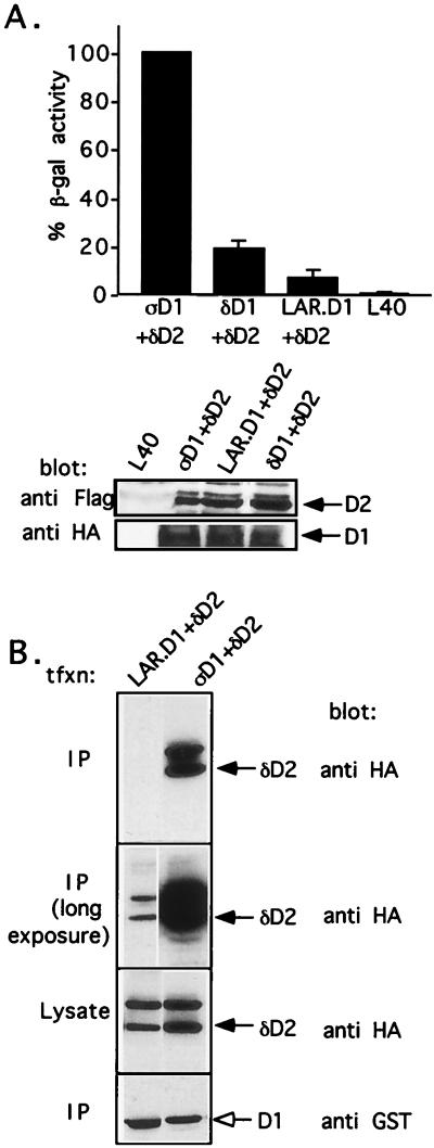
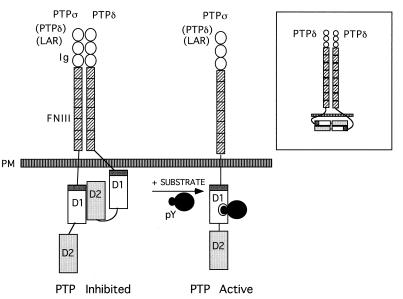
The LAR family protein tyrosine phosphatases (PTPs), including LAR, PTP delta, and PTP sigma, are transmembrane proteins composed of a cell adhesion molecule-like ectodomain and two cytoplasmic catalytic domains: active D1 and inactive D2. We performed a yeast two-hybrid screen with the first catalytic domain of PTP sigma (PTP sigma-D1) as bait to identify interacting regulatory proteins. Using this screen, we identified the second catalytic domain of PTP delta (PTP delta-D2) as an interactor of PTP sigma-D1. Both yeast two-hybrid binding assays and coprecipitation from mammalian cells revealed strong binding between PTP sigma-D1 and PTP delta-D2, an association which required the presence of the wedge sequence in PTP sigma-D1, a sequence recently shown to mediate D1-D1 homodimerization in the phosphatase RPTP alpha. This interaction was not reciprocal, as PTP delta-D1 did not bind PTP sigma-D2. Addition of a glutathione S-transferase (GST)-PTP delta-D2 fusion protein (but not GST alone) to GST-PTP sigma-D1 led to approximately 50% inhibition of the catalytic activity of PTP sigma-D1, as determined by an in vitro phosphatase assay against p-nitrophenylphosphate. A similar inhibition of PTP sigma-D1 activity was obtained with coimmunoprecipitated PTP delta-D2. Interestingly, the second catalytic domains of LAR (LAR-D2) and PTP sigma (PTP sigma-D2), very similar in sequence to PTP delta-D2, bound poorly to PTP sigma-D1. PTP delta-D1 and LAR-D1 were also able to bind PTP delta-D2, but more weakly than PTP sigma-D1, with a binding hierarchy of PTP sigma-D1 >> PTP delta-D1 > LAR-D1. These results suggest that association between PTP sigma-D1 and PTP delta-D2, possibly via receptor heterodimerization, provides a negative regulatory function and that the second catalytic domains of this and likely other receptor PTPs, which are often inactive, may function instead to regulate the activity of the first catalytic domains.


Figures

FIG. 1
FIG. 1
(A) Schematic representation of PTPς (LAR-PTP2; accession no. L11587; reference 52) and the bait used for the yeast two-hybrid screen corresponding to its first catalytic domain (PTPς-D1). Ig, Ig-like repeat; FNIII, FNIII-like repeat; TM, transmembrane domain; W, wedge motif; ςD1, PTPς-D1; ςD2, PTPς-D2. (B) Regions of PTPδ (all in the second catalytic domain, D2) isolated in yeast two-hybrid screens of rat (r) lung and mouse (m) embryo (day 11) libraries. The general architecture and domain arrangement of PTPς shown in panel A are also shared by PTPδ and LAR. Splice variants of PTPς are not shown. The asterisk refers to the mouse amino acid number, since the entire rat PTPδ has not been cloned.
FIG. 2
FIG. 2
Sequence alignment of the second catalytic domains (D2) of rat (r) and mouse (m) PTPδ, rat PTPς, and rat LAR. The signature motif is in bold letters. The arrowhead represents the starting amino acid of the smallest clone isolated, located within the conserved DYINAS sequence. The previously published frameshift in the mouse PTPδ-D2 sequence (23) has been corrected based on our own sequencing.
FIG. 3
FIG. 3
Binding of PTPς-D1 to PTPδ-D2, but not of PTPδ-D1 to PTPς-D2, analyzed by yeast two-hybrid binding assays. (A) Quantitative liquid β-gal assays (histograms) or filter β-gal assays (inset) performed with the bait PTPς-D1 (ςD1) fused to the LexA DNA binding domain and the prey PTPδ-D2 (δD2), PTPς-D2 (ςD2), or LAR D2 fused to the Gal4 transactivation domain. Interactions between D1 and D2 of PTPς were also analyzed in the opposite configuration (LexA-ςD2 and Gal4-ςD1, third bar from the left and third yeast streak clockwise from the top in the inset). The quantitative β-gal assays represent means ± standard errors of six determinations. (B) Protein expression of FLAG-tagged D2 domains of PTPς (ςD2) and PTPδ (δD2) in the pACT vector, demonstrating strong expression of both proteins in yeast L40 cells (bottom) but an association of ςD1 with δD2 only and not with ςD2 upon coexpression in L40 cells (top). (C) Lack of binding of PTPδ-D1 to PTPς-D2. Filter β-gal assays (top, inset) or liquid β-gal (top) assays were performed with the bait LexA–PTPδ-D1 and the prey Gal4–PTPς-D2 cotransformed into yeast L40 cells or with the positive control LexA–PTPς-D1 plus Gal4–PTPδ-D2. Bars represent means ± standard errors (n = 8). The actual β-gal activities were 445.7 ± 27.6 U for ςD1 plus δD2 and 6.4 ± 1.2 U for δD1 plus ςD2. For analysis of protein expression, L40 cells untransformed or transformed with δD1 plus ςD2 or with ςD1 plus δD2 were lysed and proteins were separated by SDS–10% PAGE and immunoblotted with anti-FLAG (against D2) or anti-HA (against D1) antibodies (bottom).
FIG. 4
FIG. 4
Coprecipitation of PTPς-D1 and PTPδ-D2 in mammalian cells. PTPς-D1 expressed as a GST fusion protein (in the mammalian expression vector pEBG) was transiently cotransfected with either HA-tagged PTPδ-D2, PTPς-D2, or LAR-D2 (in pCMV4) into Cos7 cells. Transfected cells were lysed, the lysate was incubated with glutathione agarose beads to precipitate GST–PTPς-D1 and associated proteins, and the proteins were separated on by SDS–10% PAGE and immunoblotted with anti-HA antibodies to detect coprecipitated HA-tagged D2 domains (panel A, IP). Aliquots of the lysate were also analyzed for levels of expression of either the HA-tagged D2 domains by using anti-HA antibodies (panel B, lysate) or GST–PTPς-D1 by using anti-GST antibodies (panel C, lysate). The blot in panel A was then stripped and reprobed with anti-GST antibodies to determine the amount of GST–PTPς-D1 precipitated from the cell lysates (panel D, IP). The reason for the appearance of a slower-migrating band (∼35 kDa, recognized by both anti-HA and anti-D2 antibodies) in lanes representing the D2 domains of PTPς, PTPδ, and LAR (B) is not known, but these bands do not represent phosphorylated forms of the proteins (data not shown). The asterisk marks the HA-tagged α subunit of the rat epithelial Na+ channel (αrENaC), which was used as a negative control for these experiments. tfxn, transfection.
FIG. 5
FIG. 5
Inhibition of catalytic activity of PTPς-D1 by PTPδ-D2. (A) Bacterially expressed GST fusion protein of PTPς-D1 (ςD1), either soluble or immobilized on glutathione agarose beads, was incubated with 100 mM PNPP either alone, with 500 ng of bacterially expressed soluble PTPδ-D2 (δD2), or with GST (control). The reaction was stopped with 0.9 ml of 1 M NaOH, and the optical density at 450 nm of the p-nitrophenolate product was measured. These data are means ± standard errors of four independent experiments. GST–PTPδ-D2 alone had no catalytic activity (data not shown). (B) Inhibition of the catalytic activity of PTPς-D1 (ςD1) by PTPδ-D2 (δD2) coprecipitated from Cos7 cells. The activity of ςD1 precipitated from Cos7 cells, transfected with either ςD1 alone or cotransfected with ςD1 plus δD2, was analyzed as described for panel A. The data are the means ± standard errors of nine independent experiments.
FIG. 6
FIG. 6
Requirement of the wedge sequence of PTPς-D1 for interaction with PTPδ-D2. (A) Alignment of the wedge (W) sequences of the first catalytic domains of several RPTPs, including mouse (m) RPTPα, rat (r) PTPς, mouse PTPδ, and rat LAR. A more detailed alignment is provided elsewhere (3). Boxed residues represent identical or conserved amino acids conforming to the consensus of the wedge sequence. The α1 and α2 helices are represented by black boxes. (B) Schematic representation of the two-hybrid baits for panel C, including full-length PTPς-D1 (ςD1) or the N-terminally truncated, wedge-deleted (ςD1-W) regions. Both proteins were expressed as fusion proteins with the LexA DNA binding domain. (C) Yeast two-hybrid binding assays of L40 cells cotrans- formed with ςD1 (bait) plus δD2 (prey), with ςD1-W (bait) plus δD2 (prey), or with each construct alone (ςD1, ςD1-W, or δD2). Quantitative β-gal assay results (histograms) are the means ± the standard errors of six determinations. Filter β-gal assay results are shown in the inset (bottom). Levels of protein expression of the PTPς-D1 and PTPςD1-W baits (HA tagged) in these experiments were similar, as determined by immunoblotting with anti-HA antibodies (top of inset, arrows). (D) Lack of coprecipitation of PTPδ-D2 with PTPςD1-W. Cos7 cells were cotransfected with HA-tagged PTPδ-D2 (δD2) together with either GST–PTPς-D1 (ςD1) or the wedge-deleted GST–PTPςD1-W (ςD1-W) construct. Cells were then lysed, and the lysate was incubated with glutathione agarose beads to precipitate GST–PTPς-D1 or GST–PTPςD1-W and associated proteins. Proteins bound to the beads were separated by SDS–10% PAGE and immunoblotted with anti-HA antibodies to test for coprecipitation of PTPδ-D2 (top panel, IP). Aliquots of the lysate of the transfected cells were analyzed for levels of expression of PTPδ-D2 by using anti-HA antibodies (second panel) or GST–PTPς-D1 and GST–PTPςD1-W by using anti-GST antibodies (third panel). The blot in the top panel was then stripped and reprobed with anti-GST antibodies to determine the amount of GST–PTPς-D1 or GST–PTPςD1-W precipitated from the transfected cell lysate (IP, bottom panel). tfxn, transfection.
FIG. 7
FIG. 7
Weak association of the D1 of other LAR family members (PTPδ and LAR) with PTPδ-D2. (A) Liquid β-gal assays performed with the bait PTPς-D1 (ςD1), PTPδ-D1 (δD1), or LAR-D1 fused to the LexA DNA binding domain either alone or together with the prey PTPδ-D2 (δD2) fused to the Gal4 transactivation domain. L40 represents the β-gal activity of untransformed yeast cells. The data are percentages of the β-gal activity of ςD1 plus δD2 and represent the means ± the standard errors of 8 to 24 determinations. One hundred percent β-gal activity of ςD1 plus δD2 corresponds to 423 ± 40 U. There was no autoactivation of each domain alone. For analysis of protein expression, L40 cells untransformed or transformed with ςD1 plus δD2, δD1 plus δD2, LAR-D1 plus δD2, or the domains alone were lysed, and the proteins were separated by SDS–10% PAGE and immunoblotted with anti-FLAG (against D2) or anti-HA (against D1) antibodies. (B) Poor association of LAR-D1 with δD2 in mammalian cells. Cos7 cells were cotransfected with GST–LAR-D1 (LAR-D1) or GST-ςD1 (ςD1) (used as a control) together with HA-tagged PTPδ-D2 (δD2). Cells were lysed, the lysate was incubated with glutathione agarose beads to precipitate the GST-tagged D1s and their associated proteins, and the proteins were separated by SDS–10% PAGE and immunoblotted with either anti-GST antibodies to detect the D1 proteins (lowest panel) or anti-HA antibodies to detect coprecipitated δD2 (upper two panels, showing short- and long-exposure autoradiograms). Equal expression of δD2 in all transfections was confirmed by immunoblotting the lysate of transfected cells with anti-HA antibodies (third panel from the top). tfxn, transfection.
FIG. 8
FIG. 8
Model of D1-D2 heterodimerization of LAR family PTPs. Under resting conditions, the first catalytic domain (D1) of PTPς (or possibly other LAR family PTPs [in parentheses]) is associated with the second catalytic domain (D2) of PTPδ, an association requiring the wedge sequence of PTPς (dark grey). Such intermolecular dimerization inhibits catalytic activity, thus keeping the phosphatase in a partially inactive state. Our current data do not support the association of PTPς-D1 with PTPς-D2 or LAR-D1 with LAR-D2 but cannot preclude the possibility of a weak inter- or intramolecular interaction of PTPδ-D1 with PTPδ-D2 (inset). We speculate that upon presentation of the as-yet-unidentified tyrosine-phosphorylated substrate (likely of high affinity), the D1-D2 heterocomplex is likely to dissociate, thus allowing substrate dephosphorylation. PM, plasma membrane.

Similar articles

Cited by

References

    1. Aicher B, Lerch M M, Müller T, Schilling J, Ullrich A. Cellular redistribution of protein tyrosine phosphatases LAR and PTPς by inducible proteolytic processing. J Cell Biol. 1997;138:681–696. - PMC - PubMed
    1. Barnea G, Silvennoinen O, Shaanan B, Honegger A M, Canoll P D, D’Eustachio P, Morse B, Levy J B, LaForgia S, Huebner K, Musacchio J M, Sap J, Schlessinger J. Identification of a carbonic anhydrase-like domain in the extracellular region of RPTPγ defines a new subfamily of receptor tyrosine phosphatases. Mol Cell Biol. 1993;13:1497–1506. - PMC - PubMed
    1. Bilwes A M, den Hertog J, Hunter T, Noel J P. Structural basis for inhibition of receptor protein-tyrosine-α phosphatase by dimerization. Nature. 1996;382:555–559. - PubMed
    1. Brady-Kalnay S M, Flint A J, Tonks N K. Homophilic binding of PTPμ, a receptor type protein tyrosine phosphatase, can mediate cell-cell aggregation. J Cell Biol. 1993;122:961–972. - PMC - PubMed
    1. Brady-Kalnay S M, Tonks N K. Identification of the homophilic binding site of the receptor protein tyrosine phosphatase PTPμ. J Biol Chem. 1994;269:28472–28477. - PubMed

Publication types

MeSH terms

Substances

LinkOut - more resources