. 1998 May;18(5):2629-39.
doi: 10.1128/MCB.18.5.2629.

Cooperative Pho2-Pho4 interactions at the PHO5 promoter are critical for binding of Pho4 to UASp1 and for efficient transactivation by Pho4 at UASp2

Affiliations

Cooperative Pho2-Pho4 interactions at the PHO5 promoter are critical for binding of Pho4 to UASp1 and for efficient transactivation by Pho4 at UASp2

S Barbaric et al. Mol Cell Biol. 1998 May.

Abstract

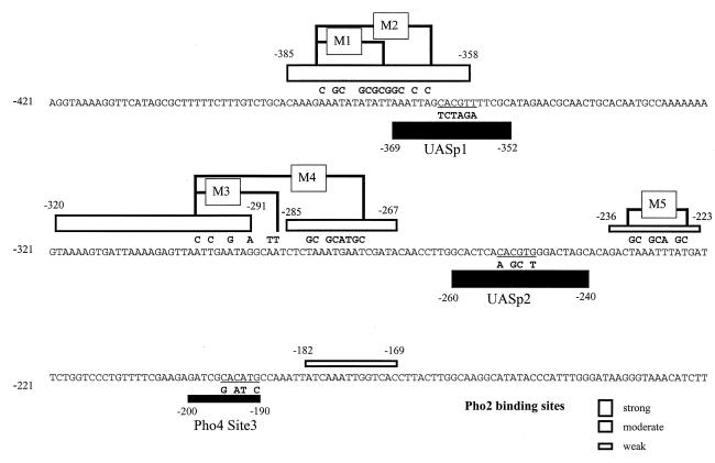
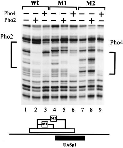
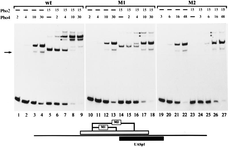
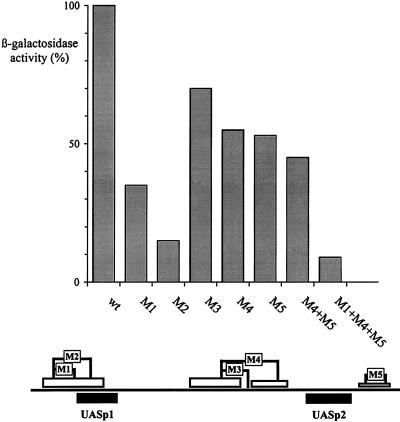
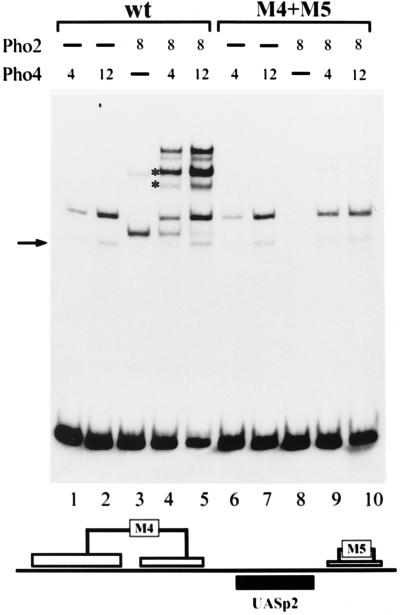
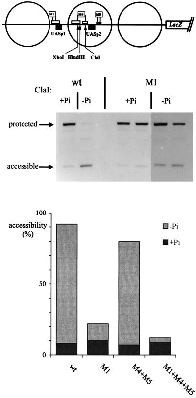
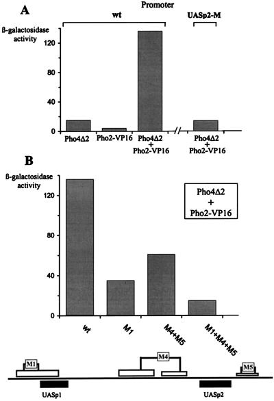
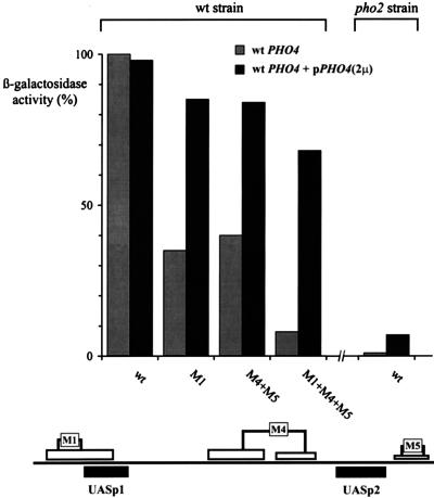
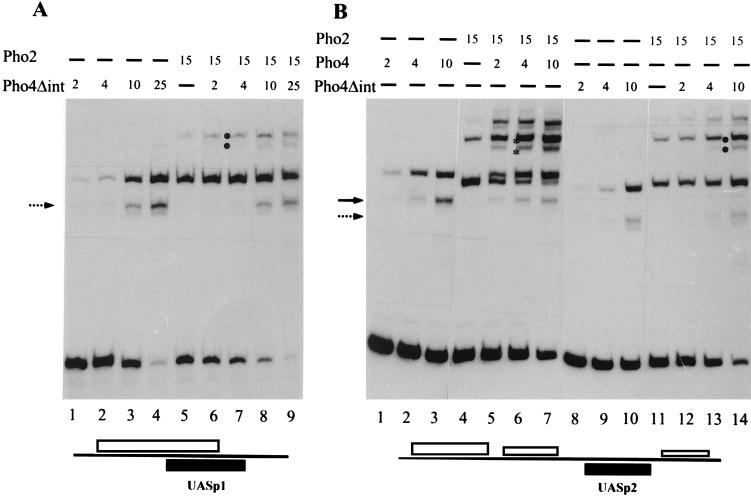
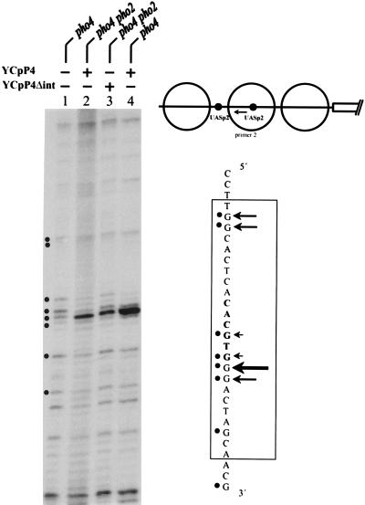
The activation of the PHO5 gene in Saccharomyces cerevisiae in response to phosphate starvation critically depends on two transcriptional activators, the basic helix-loop-helix protein Pho4 and the homeodomain protein Pho2. Pho4 acts through two essential binding sites corresponding to the regulatory elements UASp1 and UASp2. Mutation of either of them results in a 10-fold decrease in promoter activity, and mutation of both sites renders the promoter totally uninducible. The role of Pho4 appears relatively straightforward, but the mechanism of action of Pho2 had remained elusive. By in vitro footprinting, we have recently mapped multiple Pho2 binding sites adjacent to the Pho4 sites, and by mutating them individually or in combination, we now show that each of them contributes to PHO5 promoter activity. Their function is not only to recruit Pho2 to the promoter but to allow cooperative binding of Pho4 together with Pho2. Cooperativity requires DNA binding of Pho2 to its target sites and Pho2-Pho4 interactions. A Pho4 derivative lacking the Pho2 interaction domain is unable to activate the promoter, but testing of UASp1 and UASp2 individually in a minimal CYC1 promoter reveals a striking difference between the two UAS elements. UASp1 is fully inactive, presumably because the Pho4 derivative is not recruited to its binding site. In contrast, UASp2 activates strongly in a Pho2-independent manner. From in vivo footprinting experiments and activity measurements with a promoter variant containing two UASp2 elements, we conclude that at UASp2, Pho2 is mainly required for the ability of Pho4 to transactivate.


Figures

FIG. 1
FIG. 1
Mutation of Pho2 and Pho4 binding sites at the PHO5 promoter. The locations of the Pho4 and Pho2 binding sites as determined by in vitro footprinting (5) are indicated by solid and open bars, respectively, the width of the bars corresponding to the relative affinities of the sites for the factor. Mutated regions within the Pho2 binding sites are in boxes (M1 to M5), and the changed nucleotides are shown above the wild-type sequence. Mutations within the Pho4 consensus sequence are shown below the wild-type sequence and are referred to as UASp1-mut, UASp2-mut, and Pho4 Site3-mut.
FIG. 2
FIG. 2
Effect of the mutations M1 and M2 on Pho2 and Pho4 binding in vitro. DNase I footprinting was performed as described in Materials and Methods. The upper strand of an SfuI (−206)-BamHI (−542) fragment, derived from the wild-type or mutated promoter, was labeled at the SfuI site. Pho4 or Pho2 was added as indicated at the top. The regions protected in the wild-type promoter are indicated on the side. The locations of the M1 and M2 mutations (Fig. 1) within the Pho2 binding site are shown schematically underneath.
FIG. 3
FIG. 3
Pho2 DNA binding is required for cooperativity between Pho2 and Pho4 at UASp1. The binding reaction and the gel shift assay were performed as described in Materials and Methods. A labeled 81-bp PCR promoter fragment (−324 to −405) was used, containing either the wild-type promoter sequence or the M1 or M2 mutation (see Fig. 1), and is schematically shown at the bottom. The amounts of protein added to an assay mixture are indicated in arbitrary units. One unit of Pho4 and Pho2 corresponds to about 5 and 6 ng of protein, respectively, as determined by sodium dodecyl sulfate gel electrophoresis. The higher-mobility protein-DNA complex observed with Pho4 added alone (marked by an arrow) represents proteolytically degraded Pho4 protein bound to DNA. The positions of the ternary complexes containing either full-length or degraded Pho4 protein are indicated by asterisks. A lower-mobility complex with only Pho2 (lane 5) migrates at approximately the same position as the ternary complex. However, the presence of a ternary complex with the proteolyzed Pho4 protein (lower band with asterisk) makes it possible to unambiguously identify the ternary complex.
FIG. 4
FIG. 4
Mutations in the Pho2 binding sites differentially affect PHO5 promoter activity. The activities of the wild-type PHO5 promoter fused to the lacZ gene (26) and of promoter variants containing mutations in the Pho2 binding sites were measured as described in Materials and Methods. The activities of the mutated promoter variants are expressed relative to the activity of the wild-type promoter (920 U). The mutations are schematically shown at the bottom.
FIG. 5
FIG. 5
Pho2-DNA binding is required for cooperativity between Pho2 and Pho4 also at UASp2. The binding reaction and the gel shift assay were performed as described in Materials and Methods. A labeled 109-bp PCR-generated promoter fragment (−316 to −208) containing UASp2 and either the wild-type (wt) sequence or the combined M4+M5 mutation, shown schematically at the bottom, were used. The amounts of protein added to the assay mixture are listed in arbitrary units (for details, see the legend to Fig. 3). For the explanation of the arrow and the asterisks, see the legend to Fig. 3.
FIG. 6
FIG. 6
Chromatin opening at the PHO5 promoter depends on the Pho2 cis elements adjacent to UASp1 but not at UASp2. Strains carrying either the wild-type (wt) PHO5-lacZ plasmid or plasmids with promoter variants were grown in media containing Pi (+Pi) or not containing Pi (−Pi) as indicated, and nuclei were prepared. They were digested for 60 min at 37°C in 200 μl of buffer with 100 U of ClaI or 200 U of HindIII or XhoI (the M4 mutation introduces an XhoI site and a HindIII site, whereas the ClaI site is destroyed). In order to monitor cleavage by the restriction nuclease at the sites shown in the schematic at the top, DNA was isolated, cleaved with RsaI, analyzed in a 1% agarose gel, blotted, and hybridized with a pBR322 RsaI-BamHI fragment which hybridizes to the region immediately upstream of the BamHI site. A 1.46-kb RsaI fragment is generated if the restriction nuclease had been protected, and a fragment about half that size is generated if the site had been accessible. Analysis of the wild-type reporter and the M1 promoter is shown at the top. Accessibility values for the wild-type reporter and M1 promoter as well as M4+M5 and M1+M4+M5 are shown in the diagram below. Measurements for M4+M5 and M1+M4+M5 were derived from XhoI and HindIII digests which gave values that were within 5% of each other.
FIG. 7
FIG. 7
Pho2 cis elements are required for activation of the PHO5 promoter by Pho2-VP16 and a transcriptionally inactive Pho4 derivative. (A) Activation of the wild-type PHO5 promoter or a promoter variant with a mutated Pho4 site at UASp2 (UASp2-M) by Pho4Δ2 and/or Pho2-VP16 as measured in YS22 (pho2) or YS27 (pho4 pho2). (B) Activation of the PHO5 promoter variants containing mutated Pho2 binding sites, shown schematically at the bottom, by Pho4Δ2 and Pho2-VP16 expressed together in YS27 (pho4 pho2).
FIG. 8
FIG. 8
Overexpression of Pho4 relieves the requirement for Pho2 cis-acting elements. Activation of PHO5 promoter variants containing mutated Pho2 cis elements, indicated schematically at the bottom, was measured in a wild-type (wt) strain (YS18) and in the same strain expressing Pho4 from a multicopy plasmid. The activation of a wild-type reporter in a pho2 strain (YS19) is shown on the right. 2μ, 2μm plasmid.
FIG. 9
FIG. 9
Cooperative DNA binding with Pho2 is largely abolished with a Pho4 variant lacking the Pho2 interaction domain. The binding reaction and the gel shift assay were performed as described in Materials and Methods. Proteins were added individually or in combination in the amounts indicated (arbitrary units as in Fig. 3, with 1 U of Pho4Δint corresponding to about 5 ng of protein) to a labeled 81-bp PCR-generated promoter fragment (−324 to −405) containing UASp1 and the overlapping Pho2 sites (A), or to a labeled 109-bp PCR-generated fragment (−316 to −208) containing UASp2 and the adjacent Pho2 sites (B) (see schematics at the bottom). The arrows mark binary complexes derived from proteolytically degraded Pho4 (solid arrow) and Pho4Δint (broken arrow). Ternary complexes with full-length Pho4 and its degradation product are marked by asterisks and those for Pho4Δint are marked analogously by dots.
FIG. 10
FIG. 10
DMS footprint analysis of Pho4 binding to UASp2. Binding of wild-type Pho4 and Pho4Δint to the upstream UASp2 element in the PHO5 promoter variant in which UASp1 was replaced by UASp2 (see schematic) was examined in YS22 (pho4) or YS27 (pho2 pho4). For details of DMS footprinting and the primer used (arrow in the schematic), see Materials and Methods.

References

    1. Almer A, Hörz W. Nuclease hypersensitive regions with adjacent positioned nucleosomes mark the gene boundaries of the PHO5/PHO3 locus in yeast. EMBO J. 1986;5:2681–2687. - PMC - PubMed
    1. Almer A, Rudolph H, Hinnen A, Hörz W. Removal of positioned nucleosomes from the yeast PHO5 promoter upon PHO5 induction releases additional upstream activating DNA elements. EMBO J. 1986;5:2689–2696. - PMC - PubMed
    1. Arndt K T, Styles C, Fink G R. Multiple global regulators control HIS4 transcription in yeast. Science. 1987;237:874–880. - PubMed
    1. Barbaric S, Fascher K D, Hörz W. Activation of the weakly regulated PHO8 promoter in S. cerevisiae: chromatin transition and binding sites for the positive regulator protein PHO4. Nucleic Acids Res. 1992;20:1031–1038. - PMC - PubMed
    1. Barbaric S, Münsterkötter M, Svaren J, Hörz W. The homeodomain protein Pho2 and the basic-helix-loop-helix protein Pho4 bind DNA cooperatively at the yeast PHO5 promoter. Nucleic Acids Res. 1996;24:4479–4486. - PMC - PubMed

Publication types