. 1998 May;18(5):2729-37.
doi: 10.1128/MCB.18.5.2729.

c-Maf interacts with c-Myb to regulate transcription of an early myeloid gene during differentiation

Affiliations

c-Maf interacts with c-Myb to regulate transcription of an early myeloid gene during differentiation

S P Hedge et al. Mol Cell Biol. 1998 May.

Abstract

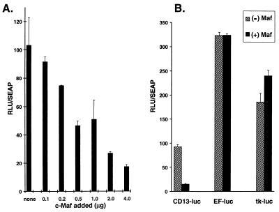
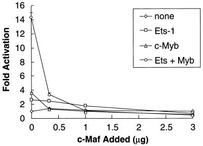
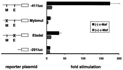
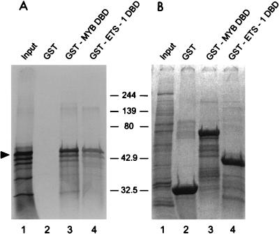
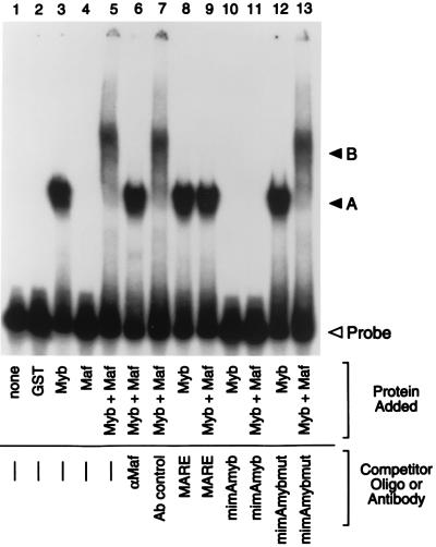
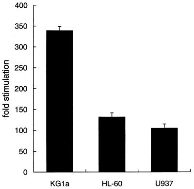
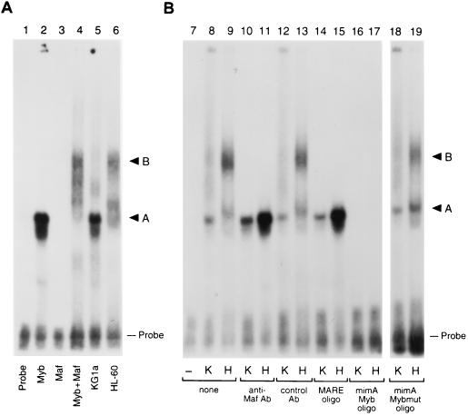
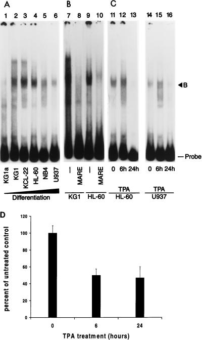
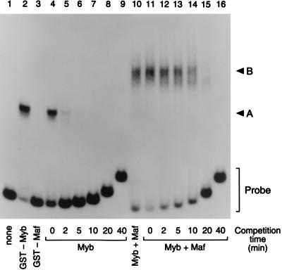
The MafB transcriptional activator plays a pivotal role in regulating lineage-specific gene expression during hematopoiesis by repressing Ets-1-mediated transcription of key erythroid-specific genes in myeloid cells. To determine the effects of Maf family proteins on the transactivation of myeloid-specific genes in myeloid cells, we tested the ability of c-Maf to influence Ets-1- and c-Myb-dependent CD13/APN transcription. Expression of c-Maf in human immature myeloblastic cells inhibited CD13/APN-driven reporter gene activity (85 to 95% reduction) and required the binding of both c-Myb and Ets, but not Maf, to the promoter fragment. c-Maf's inhibition of CD13/APN expression correlates with its ability to physically associate with c-Myb. While c-Maf mRNA and protein levels remain constant during myeloid differentiation, formation of inhibitory Myb-Maf complexes was developmentally regulated, with their levels being highest in immature myeloid cell lines and markedly decreased in cell lines representing later developmental stages. This pattern matched that of CD13/APN reporter gene expression, indicating that Maf modulation of c-Myb activity may be an important mechanism for the control of gene transcription during hematopoietic cell development.


Figures

FIG. 1
FIG. 1
c-Maf selectively abrogates transcription from CD13/APN promoter constructs in myeloid cells. (A) c-Maf inhibition is dose dependent. Increasing amounts (0.1 to 4.0 μg) of the expression construct pRc/RSVcMaf were cotransfected with 4 μg of the CD13/APN wild-type reporter construct (−411luc) into KG1a immature myeloblastic cells, and luciferase activity was assayed at 24 h. (B) Inhibition by c-Maf is selective. The indicated reporter plasmids (4 μg) were transfected with equal amounts of c-Maf expression plasmids or the empty expression vector. All values are normalized to those for a cotransfected control plasmid (MAP1-SEAP). RLU, relative light units.
FIG. 2
FIG. 2
c-Maf affects the c-Myb–Ets-1 functional cooperation on the CD13/APN promoter. The C33A human epithelial cell line was cotransfected with 1 μg of the −411luc reporter construct and increasing amounts (0.3 to 3.0 μg) of pRc/RSVcMaf along with either empty vectors only (none), 0.5 μg of pEVRFO-Ets-1 only (Ets-1), 1 μg of pCMV4cMyb only (c-Myb), or 1 μg of pCMV4cMyb cotransfected with 0.5 μg of pEVRFO-Ets-1 (Ets-1 + Myb). All values are normalized to those for a cotransfected control plasmid (MAP1-SEAP) both for transfection efficiency and as a control for the c-Myb, Ets, and Maf effect on other promoters. Results are expressed as fold activation above that obtained with cotransfection of equal amounts of the empty expression plasmid.
FIG. 3
FIG. 3
c-Maf inhibits c-Myb and Ets-1 transcriptional activity. Representations of the wild-type reporter plasmid (−411luc) indicating the c-Myb and Ets-1 consensus sites, and those containing deletions from the 5′ end (−291luc), point mutations (Mybmut), or internal deletions (Etsdel) of the CD13/APN myeloid promoter, are depicted on the left. Reporter plasmids (5 μg) were transiently transfected into the immature myeloblastic cell line KG1a along with 5 μg of pRc/RSVcMaf expression construct [(+) c-Maf)] or the empty expression vector [(−) c-Maf] and assayed for luciferase activity. All data are normalized to values for the cotransfected MAP1-SEAP control plasmid both for transfection efficiency and as a control for c-Maf’s effect on other promoters. Results are expressed as fold stimulation above the normalized activity of the empty reporter plasmid pGL2basic.
FIG. 4
FIG. 4
Full-length c-Maf binds to the c-Myb and Ets-1 DNA binding domains. (A) [35S]methionine-labeled c-Maf produced in vitro was incubated with GST alone (lanes 2) or with GST-Myb-DBD (amino acids 1 to 240) (lane 3) or GST-Ets-1-DBD (amino acids 322 to 440) (lane 4) fusion protein. The interacting proteins were purified on GSH-beads and analyzed by SDS-PAGE and autoradiography. The arrowhead indicates full-length c-Maf; molecular size markers are indicated in kilodaltons. Input (lane 1) shows 10% of the c-Maf used in each pull-down assay. GST acts as a negative control for c-Maf binding. (B) Coomassie blue staining of the same gel indicates that comparable amounts of GST fusion proteins were used in each assay.
FIG. 5
FIG. 5
c-Myb and Maf proteins form higher-order complexes on the CD13/APN promoter in vitro. An end-labeled 135-bp promoter fragment probe (bp −426 through −291) containing the functionally defined Maf target sequences was incubated with bacterially expressed GST fusion proteins. Lane 1, probe alone; lane 2, GST control; lanes 3, 8, 10, and 12, GST-Myb-DBD only; lane 4, GST-Maf only; lanes 5 to 7, 9, 11, and 13, equal amounts of GST-Myb-DBD and GST-Maf. Antibodies recognizing the c-Maf protein (lane 6) or an isotype-matched control antibody (Ab) (lane 7) were added in equal amounts to binding reactions. Unlabeled competitor oligonucleotides containing either the consensus Maf binding site (MARE; lanes 8 and 9), the consensus Myb site from the mim-1 promoter (mimAmyb; lanes 10 and 11), or a mutated Myb site (mimAmybmut; lanes 12 and 13) were added to the assays in 100-fold molar excess. A, the specific DNA-protein complex formed by GST-Myb-DBD; B, the ternary complex containing DNA, c-Myb, and c-Maf; Probe, free probe.
FIG. 6
FIG. 6
The activity of CD13/APN promoter constructs is diminished in later-stage myeloid cells. The −411luc reporter construct containing sequences sufficient for wild-type level, tissue-appropriate expression from the CD13/APN myeloid promoter (60) was transiently transfected into the immature myeloblastic cell line KG1a, the promyelocytic cell line HL-60, or the monoblastic cell line U937 and assayed for luciferase activity at 6 h. Luciferase activities were normalized for differences in transfection efficiency between cell lines with SEAP activity produced by the cotransfected plasmid MAP1-SEAP. Results are expressed as fold stimulation above the normalized activity of the empty reporter plasmid pGL2basic in each cell line.
FIG. 7
FIG. 7
Expression of c-Maf and c-Myb in myeloid cell lines. (A) c-Maf mRNA expression levels in human myeloid cell lines as determined by Northern blot analysis of poly(A)+ RNA from the KG1a immature myeloblastic cell line or the HL-60 promyelocytic cell line. A single blot was sequentially probed, stripped, and reprobed with the β-actin probe as a control for RNA loading and integrity. Exposure times for optimal detection of c-Maf were routinely significantly longer than for β-actin (1 week for c-Maf versus 1 day for β-actin). (B) c-Maf protein levels in human myeloid cell lines as determined by immunoprecipitation followed by Western blot analysis. Cell lysates from the KG1a and HL-60 cell lines were immunoprecipitated with either an antiserum recognizing the c-Maf protein (lanes 3 and 4) or an isotype-matched control (lanes 5 and 6). Immunoprecipitates were analyzed beside control lysates from an induced (lane 7) or uninduced (lane 8) cell line engineered to inducibly express a full-length c-Maf expression construct or IgG protein alone (lane 9). The gel was transferred and probed for c-Maf protein with the anti-Maf antiserum. The arrowhead marks the specific c-Maf band; asterisks indicate IgG bands recognized by the secondary donkey anti-rabbit IgG antiserum.
FIG. 8
FIG. 8
Myb-Maf complex formation is regulated differently in early- and later-stage myeloid cell lines. An end-labeled 135-bp promoter fragment probe containing the functionally defined Maf target sequences was incubated with bacterially expressed GST fusion proteins or whole-cell lysates from KG1a immature myeloblasts or HL-60 promyelocytes. (A) HL-60 lysates contain a complex that comigrates with the Myb-Maf complex (GST-Myb-DBD–GST-Maf complex, 57 kDa + 69 kDa = 126 kDa; native Myb-Maf complex, 80 kDa + 42 kDa = 122 kDa). Lane 1, probe alone; lane 2, GST-Myb-DBD only; lane 3, GST-Maf only; lane 4, equal amounts of GST-Myb-DBD and GST-Maf; lane 5, KG1a lysate; lane 6, HL-60 lysate. (B) HL-60 lysates contain a Myb-Maf complex that binds to the Myb site. Lane 7, probe alone; lanes 8, 10, 12, 14, 16, and 18, KG1a lysate (K); lanes 9, 11, 13, 15, 17, and 19, HL-60 lysate (H). Antibodies (Ab) recognizing the c-Maf protein (lanes 10 and 11) or an isotype-matched control antibody (lanes 12 and 13) were added in equal amounts to binding reactions. Unlabeled competitor oligonucleotides containing either the consensus Maf binding site (MARE; lanes 14 and 15), the consensus Myb site from the mim-1 promoter (mimAmyb; lanes 16 and 17), or a mutated Myb site (mimAmybmut; lanes 18 and 19) were added to the assays in 100-fold molar excess. A, the DNA-protein complex containing Myb; B, the ternary complex containing both Myb and Maf; Probe, uncomplexed probe.
FIG. 9
FIG. 9
Levels of Myb-Maf complexes change with differentiation stage of myeloid cell lines and in response to monocytic differentiation signals and correlate with CD13/APN transcriptional activity. An end-labeled 135-bp promoter fragment probe containing the functionally defined Maf target sequences was incubated with whole-cell lysates from myeloid cell lines arrested at progressively more differentiated stages (37, 51) or from cell lines treated with TPA. (A) Myb-Maf complex formation appears to peak near the late myeloblast stage. Lane 1, KG1a (phenotypically primitive, developmentally arrested revertant of the KG1 cell line); lane 2, KG1 (early myeloblastic); lane 3, KCL22 (late myeloblastic); lane 4, HL-60 (promyelocytic); lane 5, NB4 (promyelocytic); lane 6, U937 (myelomonoblastic). (B) The differentially regulated complex contains Maf. Unlabeled competitor oligonucleotides containing the consensus Maf binding site (MARE; lanes 8 and 10) were added to binding reactions containing KG1 or HL-60 lysates in 100-fold molar excess. (C) Myb-Maf complexes are regulated during monocytic differentiation. HL-60 (lanes 11 to 13) or U937 (lanes 14 to 16) cells were treated with 10−6 M TPA and harvested at the indicated times. B, the secondary complex containing both Myb and Maf; Probe, uncomplexed probe. (D) Reporter gene levels correlate with complex formation. HL-60 cells were transiently transfected with 4 μg of the CD13/APN wild-type reporter construct (−411luc). Cultures were treated with 10−6 M TPA, and luciferase activity was assayed at 6 and 24 h.
FIG. 10
FIG. 10
Myb-Maf complexes are more stable than Myb alone. Mobility shift assays measuring the off rate of c-Myb alone (lanes 4 to 9) and the Myb-Maf protein complex (lanes 11 to 16) from preformed DNA-protein complexes after incubation for the indicated times with an excess (50 ng) of unlabeled competitor DNA (Myb/Ets-70; see Materials and Methods). An end-labeled 135-bp promoter fragment probe (bp −426 through −291) was incubated with bacterially expressed GST fusion proteins. Lane 1, probe alone; lanes 2 and 4 to 9, GST-Myb-DBD only; lane 3, GST-Maf only; lanes 10 to 16, equal amounts of GST-Myb-DBD and GST-Maf. A, the specific protein complex formed by GST-Myb-DBD; B, the ternary complex containing both Myb and Maf; Probe, free probe.

References

    1. Bram R J, Hung D T, Martin P K, Schreiber S L, Crabtree G R. Identification of the immunophilins capable of mediating inhibition of signal transduction by cyclosporin A and FK506: roles of calcineurin binding and cellular location. Mol Cell Biol. 1993;13:4760–4769. - PMC - PubMed
    1. Brelvi Z S, Studzinski G. Coordinate expression of c-myc, c-myb, and histone H4 genes in reversibly differentiating HL-60 cells. J Cell Physiol. 1987;131:43–49. - PubMed
    1. Burk O, Mink S, Ringwald M, Klempnauer K H. Synergistic activation of the chicken mim-1 gene by v-myb and C/EBP transcription factors. EMBO J. 1993;12:2027–2038. - PMC - PubMed
    1. Calkhoven C F, Ab G. Multiple steps in the regulation of transcription-factor level and activity. Biochem J. 1996;317:329–342. - PMC - PubMed
    1. Cordes S P, Barsh G S. The mouse segmentation gene kr encodes a novel basic domain-leucine zipper transcription factor. Cell. 1994;79:1025–1034. - PubMed

Publication types

MeSH terms

LinkOut - more resources