. 1998 May;18(5):3021-33.
doi: 10.1128/MCB.18.5.3021.

A promoter region mutation affecting replication of the Tetrahymena ribosomal DNA minichromosome

Affiliations

A promoter region mutation affecting replication of the Tetrahymena ribosomal DNA minichromosome

R C Gallagher et al. Mol Cell Biol. 1998 May.

Abstract

In the ciliated protozoan Tetrahymena thermophila the ribosomal DNA (rDNA) minichromosome replicates partially under cell cycle control and is also subject to a copy number control mechanism. The relationship between rDNA replication and rRNA gene transcription was investigated by the analysis of replication, transcription, and DNA-protein interactions in a mutant rDNA, the rmm3 rDNA. The rmm3 (for rDNA maturation or maintenance mutant 3) rDNA contains a single-base deletion in the rRNA promoter region, in a phylogenetically conserved sequence element that is repeated in the replication origin region of the rDNA minichromosome. The multicopy rmm3 rDNA minichromosome has a maintenance defect in the presence of a competing rDNA allele in heterozygous cells. No difference in the level of rRNA transcription was found between wild-type and rmm3 strains. However, rmm3 rDNA replicating intermediates exhibited an enhanced pause in the region of the replication origin, roughly 750 bp upstream from the rmm3 mutation. In footprinting of isolated nuclei, the rmm3 rDNA lacked the wild-type dimethyl sulfate (DMS) footprint in the promoter region adjacent to the base change. In addition, a DMS footprint in the origin region was lost in the rmm3 rDNA minichromosome. This is the first reported correlation in this system between an rDNA minichromosome maintenance defect and an altered footprint in the origin region. Our results suggest that a promoter region mutation can affect replication without detectably affecting transcription. We propose a model in which interactions between promoter and origin region complexes facilitate replication and maintenance of the Tetrahymena rDNA minichromosome.


Figures

FIG. 1
FIG. 1
Organization of regions of the Tetrahymena macronuclear rDNA minichromosome. (A) Structure of the Tetrahymena macronuclear rDNA minichromosome. One half of the palindromic 21-kb rDNA minichromosome is shown. Each half carries a single 35S rRNA transcription unit (open rectangle) containing the 17S, 5.8S, and 26S rRNA coding sequences. Each half also contains an origin of DNA replication (ORI) within the 5′ NTS (open oval). Heavy black lines, 5′ and 3′ NTSs of the rRNA genes; vertical dashed line, center of the molecule; thin black line: telomeric DNA; bent arrow, start site of rRNA transcription. (B) rDNA maintenance mutant base changes within the 1.9-kb 5′ NTS. The ∼400-bp repeats 1 and 2 are indicated. Filled rectangles, type I elements (Ia to Id). Positions of base changes are indicated both for the previously identified mutations rmm1, -3, and -4 and for the mutations identified in this work, rmm7 and -8. Sequences of type Ia, Ib, and Ic repeats: TTTTTTTGGCAAAAAAAAAAACAAAAATAGTAA; sequence of type Id repeat: ATTCTTTGGCAAAAAAAATAAAAATAATATCAG/GG (the slash in Id indicates the 3′ boundary of this repeat). Mutated residues and repeats are in boldface and are as follows: rmm1 and -4, −A in type Ib (−720); rmm3, −A in type Ic (−100); rmm7, +A in type Ib (−720); rmm8, G to A 2 nt downstream of type Id (−19) (numbers in parentheses show the distance of the mutation from the start site of transcription [approximate for rmm1, -3, -4, and -7]). (C) DNase I footprint near the start site of rRNA transcription in wild-type C3 cells. Lanes: +, footprinting of rDNA in isolated nuclei; −, control footprinting performed in parallel with purified, deproteinized T. thermophila DNA; A and T, DNA sequencing lanes. Final DNase I concentrations (units per milliliter) are shown above the lanes; nucleotide positions in the rDNA (numbered outward from center of the molecule, starting at 1 as indicated) are shown at right. Bent arrow, transcription start site at position 1887.
FIG. 1
FIG. 1
Organization of regions of the Tetrahymena macronuclear rDNA minichromosome. (A) Structure of the Tetrahymena macronuclear rDNA minichromosome. One half of the palindromic 21-kb rDNA minichromosome is shown. Each half carries a single 35S rRNA transcription unit (open rectangle) containing the 17S, 5.8S, and 26S rRNA coding sequences. Each half also contains an origin of DNA replication (ORI) within the 5′ NTS (open oval). Heavy black lines, 5′ and 3′ NTSs of the rRNA genes; vertical dashed line, center of the molecule; thin black line: telomeric DNA; bent arrow, start site of rRNA transcription. (B) rDNA maintenance mutant base changes within the 1.9-kb 5′ NTS. The ∼400-bp repeats 1 and 2 are indicated. Filled rectangles, type I elements (Ia to Id). Positions of base changes are indicated both for the previously identified mutations rmm1, -3, and -4 and for the mutations identified in this work, rmm7 and -8. Sequences of type Ia, Ib, and Ic repeats: TTTTTTTGGCAAAAAAAAAAACAAAAATAGTAA; sequence of type Id repeat: ATTCTTTGGCAAAAAAAATAAAAATAATATCAG/GG (the slash in Id indicates the 3′ boundary of this repeat). Mutated residues and repeats are in boldface and are as follows: rmm1 and -4, −A in type Ib (−720); rmm3, −A in type Ic (−100); rmm7, +A in type Ib (−720); rmm8, G to A 2 nt downstream of type Id (−19) (numbers in parentheses show the distance of the mutation from the start site of transcription [approximate for rmm1, -3, -4, and -7]). (C) DNase I footprint near the start site of rRNA transcription in wild-type C3 cells. Lanes: +, footprinting of rDNA in isolated nuclei; −, control footprinting performed in parallel with purified, deproteinized T. thermophila DNA; A and T, DNA sequencing lanes. Final DNase I concentrations (units per milliliter) are shown above the lanes; nucleotide positions in the rDNA (numbered outward from center of the molecule, starting at 1 as indicated) are shown at right. Bent arrow, transcription start site at position 1887.
FIG. 2
FIG. 2
An in vivo competition assay demonstrates the maintenance defect of rmm3 rDNA. Wild-type C3 (□) or C3-rmm3 (◊) cells were crossed with B strain cells. Progeny were maintained in log-phase growth, and total DNA was isolated every six to seven generations, cut with SphI, and Southern blotted. The graph shows the percentage of C3 rDNA as a function of increasing number of generations.
FIG. 3
FIG. 3
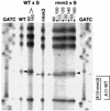
Loss of the rmm3 −A base change in the population of recombinant rDNA minichromosomes that have lost their maintenance defect. The rRNA promoter region of the rDNA minichromosome was amplified from total cell DNA isolated from each strain or progeny cell population. The C nucleotide sequencing reaction at the type Ic repeat region is shown for each sample or time point. Lanes: WT and rmm3, C sequencing reactions of wild-type C3 and rmm3 homozygotes respectively; rmm3 × B, progeny of the same B/C3-rmm3 cross shown in Fig. 2, from generations 60 to 116, as indicated above the lanes; WT × B, DNA from progeny of the B/C3 wild-type cross shown in Fig. 2 (generations 60 and 116 only); GATC, sequencing ladders of rDNA plasmid DNA. C3 rDNA has a run of 11 A residues between two C residues in the type Ic repeat; the C3-rmm3 mutant rDNA has only 10 A residues (side brackets). Hence, the next C residue in rmm3 rDNA (open arrowheads) is displaced downward by 1 nt relative to that in wild type rDNA (filled arrowheads).
FIG. 4
FIG. 4
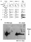
2D gel electrophoretic analysis of rDNA replicating intermediates. (A) Neutral-neutral 2D gel patterns of representative restriction fragments containing replicating DNA intermediates (3). Bubble, origin located in the center of the fragment; Simple Y, fragment passively replicated by a fork originating outside; Bubble to Y, replication bubble located asymmetrically within the fragment. Arrows indicate the directions of DNA migration. 1n, unreplicated bulk DNA fragment; 2n, almost fully replicated DNA fragment. Replicating molecules depicted below each panel run above the arc of linear DNA (connects 1n to 2n) (not illustrated here). Each number along an arc identifies the replicating molecule that runs at that position. Fork Pause, a fork pause leads to the accumulation and hence overrepresentation of a particular replicating intermediate, resulting in increased hybridization at a spot on the arc of replicating molecules (black dots 4 and 5) (see panel B). Vertical line across dark replicating intermediates, location of the fork pause. (B) Enhanced accumulation of rDNA replicating intermediates at a specific pause site in the 5′ NTS of C3-rmm3 cells. DNA from log-phase C3 wild-type or C3-rmm3 cells was restricted with HindIII. Neutral-neutral 2D gels were run as described in Materials and Methods. Double arrowhead, promoter pause (dot 5 in panel A); arrowhead, repeat 2 pause (dot 4 in panel A).
FIG. 5
FIG. 5
No effects on rRNA transcription are detected in rmm3 cells. (A) Map of rDNA minichromosome showing locations of the PCR-generated DNA probes used to analyze run-on transcription in wild-type and C3-rmm3 maintenance mutant strains; one half of the palindromic minichromosome is shown. Thin line at the end of the 3′ NTS, telomere. The expanded view of the 5′ NTS (symbols are as in Fig. 1) shows the location of the rmm3 mutation in the promoter-distal type Ic repeat. Probes: US (upstream probe), IF, ETS, 17S rRNA, and 26S rRNA. Bent arrowhead, start site of rRNA transcription; IF?, see text and panel C. (B) rmm3 homozygotes are not defective in the initiation of transcription. Run-on assays were performed on cell ghosts prepared from log-phase cells. DNA probes were those in panel A and the 5S rRNA gene, gamma tubulin gene (TUB), and lambda phage DNA. wt, wild type. (C) “IrFrt” transcripts are not altered in C3-rmm3 cells. A Northern blot of total cell RNA prepared from log-phase wild-type C3 (wt) and C3-rmm3 cells, probed with primer 12, is shown L, 100-bp ladder.
FIG. 6
FIG. 6
Summary of DMS footprinting of rDNA in isolated nuclei. (A) Map of the 5′-NTS regions in the rDNA shown footprinted in panels B and C and Fig. 7 and schematic summary of the footprinting results. Smaller bracket, region of the promoter footprint; larger bracket, region of the origin region footprint; circles, positioned nucleosomes (Nuc 1 to 7) of the 5′ NTS of the rDNA (15, 36); filled rectangles, type I elements (a, b, c, and d); open rectangles, type III elements (a to f), the sites of action of topoisomerase I (2); bent arrow, start site of rRNA transcription; arrowheads, positions of the footprinted residues shown in panels B and C and in Fig. 7 and 8 (topoisomerase I [Topo I], Nuc 5, and nt 701 and 1132). (B) DMS promoter footprint in wild-type C3 rDNA. Naked DNA, or DNA in chromatin of isolated nuclei, from wild-type C3 cells was treated with 10 mM DMS for 8 min. Treated DNA was extended with primer 12. Lanes: −, DMS-treated naked DNA; +, DMS-treated chromatin. rDNA nucleotide numbers are indicated on the side; type Ic and Id elements are bracketed. Nucleotides with enhanced DMS reactivity in chromatin are indicated by arrows. (C) C3-rmm3 lacks the wild-type DMS promoter footprint. Naked DNA, or DNA in chromatin of isolated nuclei, from C3-rmm3 maintenance mutant cells was treated with 10 mM DMS for 2 min (the same patterns were obtained by treatment for 8 min [data not shown]). All else was as in panel B above.
FIG. 6
FIG. 6
Summary of DMS footprinting of rDNA in isolated nuclei. (A) Map of the 5′-NTS regions in the rDNA shown footprinted in panels B and C and Fig. 7 and schematic summary of the footprinting results. Smaller bracket, region of the promoter footprint; larger bracket, region of the origin region footprint; circles, positioned nucleosomes (Nuc 1 to 7) of the 5′ NTS of the rDNA (15, 36); filled rectangles, type I elements (a, b, c, and d); open rectangles, type III elements (a to f), the sites of action of topoisomerase I (2); bent arrow, start site of rRNA transcription; arrowheads, positions of the footprinted residues shown in panels B and C and in Fig. 7 and 8 (topoisomerase I [Topo I], Nuc 5, and nt 701 and 1132). (B) DMS promoter footprint in wild-type C3 rDNA. Naked DNA, or DNA in chromatin of isolated nuclei, from wild-type C3 cells was treated with 10 mM DMS for 8 min. Treated DNA was extended with primer 12. Lanes: −, DMS-treated naked DNA; +, DMS-treated chromatin. rDNA nucleotide numbers are indicated on the side; type Ic and Id elements are bracketed. Nucleotides with enhanced DMS reactivity in chromatin are indicated by arrows. (C) C3-rmm3 lacks the wild-type DMS promoter footprint. Naked DNA, or DNA in chromatin of isolated nuclei, from C3-rmm3 maintenance mutant cells was treated with 10 mM DMS for 2 min (the same patterns were obtained by treatment for 8 min [data not shown]). All else was as in panel B above.
FIG. 7
FIG. 7
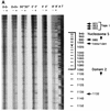
DMS-reactive A at residue position 1132 in domain 2 (in repeat 2) of C3 wild-type rDNA but not in rmm3 mutant rDNA. (A) DMS footprinting of wild-type C3 rDNA. Chromatin in nuclei isolated from wild-type C3 strain cells was footprinted with DMS. Samples were treated for 0 or 30 s or 2, 4, or 8 min, as indicated. Lanes: −, naked DNA; +, chromatin. The 30-s (+) timepoint was underloaded. The footprinted region shown extends from just upstream of the type Ib repeat towards the center of the molecule, through a highly positioned nucleosome, labeled Nucleosome 5 (see Fig. 6A). The position of nucleosome 5 is bracketed. The asterisk marks its center. Nucleotide numbers are on the side. (B) C3-rmm3 rDNA has no DMS-reactive A at nucleotide 1132 in domain 2. Chromatin from C3-rmm3 maintenance mutant cells was footprinted with DMS as described for panel A. The footprinted region is as described for panel A.
FIG. 7
FIG. 7
DMS-reactive A at residue position 1132 in domain 2 (in repeat 2) of C3 wild-type rDNA but not in rmm3 mutant rDNA. (A) DMS footprinting of wild-type C3 rDNA. Chromatin in nuclei isolated from wild-type C3 strain cells was footprinted with DMS. Samples were treated for 0 or 30 s or 2, 4, or 8 min, as indicated. Lanes: −, naked DNA; +, chromatin. The 30-s (+) timepoint was underloaded. The footprinted region shown extends from just upstream of the type Ib repeat towards the center of the molecule, through a highly positioned nucleosome, labeled Nucleosome 5 (see Fig. 6A). The position of nucleosome 5 is bracketed. The asterisk marks its center. Nucleotide numbers are on the side. (B) C3-rmm3 rDNA has no DMS-reactive A at nucleotide 1132 in domain 2. Chromatin from C3-rmm3 maintenance mutant cells was footprinted with DMS as described for panel A. The footprinted region is as described for panel A.
FIG. 8
FIG. 8
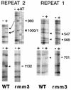
DMS reactivity of A residues in repeats 1 and 2 in wild-type (WT) and rmm3 nuclei. (Top panels) DMS-reactive A residues (filled arrowheads) in nucleosome 5 (repeat 2) and nucleosome 4 (repeat 1) have similar reactivities in wild-type and rmm3 nuclei. (Bottom panels) The A residues at corresponding positions (nt 1132 and 701) in repeats 2 and 1, respectively, are DMS reactive in wild-type nuclei (filled arrowheads) but not in rmm3 nuclei (open arrowheads). For all reactions, marker A and T sequencing reactions were run next to the lanes shown (shown only for rmm3 repeat 2 nucleosome 5 region). Footprinting was performed as described for Fig. 7, in separate experiments, and with DNA and nuclear preparations different from those shown in Fig. 7.
FIG. 9
FIG. 9
Proposed protein-DNA interactions in the rDNA 5′ NTS and promoter. (A) Map of the rDNA 5′ NTS and promoter region. Numbering of nucleotides is indicated at the rDNA center and transcription start site (1 and 1887). Nucleosomal protections (small circles) and the promoter region footprint (oval), described in this and other work (15, 36, 39) are indicated. The large circles indicate putative protein complexes at the nonnucleosomal domains 1 and 2 of repeats 1 and 2, respectively. Arrowheads demonstrate the positions of the DMS-reactive A residues identified in this work in domains 1 and 2 (positions 701 and 1132), whose reactivity is lost in rmm3 rDNA. The position of the rmm3 mutation (−A) is indicated. (B) Model of potential interactions between complexes at the promoter and upstream regions. Cylinders, highly positioned nucleosomes that package most of the DNA of the 5′ NTS (15); circles, DNA replication origin recognition factors bound to the DNA; oval, factors bound at the rRNA promoter.

Similar articles

Cited by

References

    1. Blackburn, E., D. Larson, and E. Orias. Unpublished data.
    1. Bonven B J, Gocke E, Westergaard O. A high affinity topoisomerase I binding sequence is clustered at DNAase I hypersensitive sites in Tetrahymena R-chromatin. Cell. 1985;41:541–551. - PubMed
    1. Brewer B J, Fangman W L. The localization of replication origins on ARS plasmids in S. cerevisiae. Cell. 1987;51:463–471. - PubMed
    1. Brown T. Current protocols in molecular biology. New York, N.Y: John Wiley & Sons; 1993. pp. 2.9.1–2.9.6.
    1. Budarf M L, Blackburn E H. Chromatin structure of the telomeric region and 3′-nontranscribed spacer of Tetrahymena ribosomal RNA genes. J Biol Chem. 1986;261:363–369. - PubMed

Publication types

LinkOut - more resources