. 1998 May 15;18(10):3699-707.
doi: 10.1523/JNEUROSCI.18-10-03699.1998.

Upregulation of pleiotrophin gene expression in developing microvasculature, macrophages, and astrocytes after acute ischemic brain injury

Affiliations

Upregulation of pleiotrophin gene expression in developing microvasculature, macrophages, and astrocytes after acute ischemic brain injury

H J Yeh et al. J Neurosci. .

Abstract

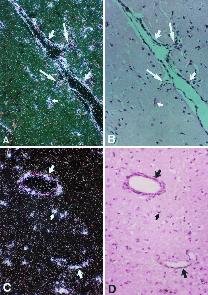
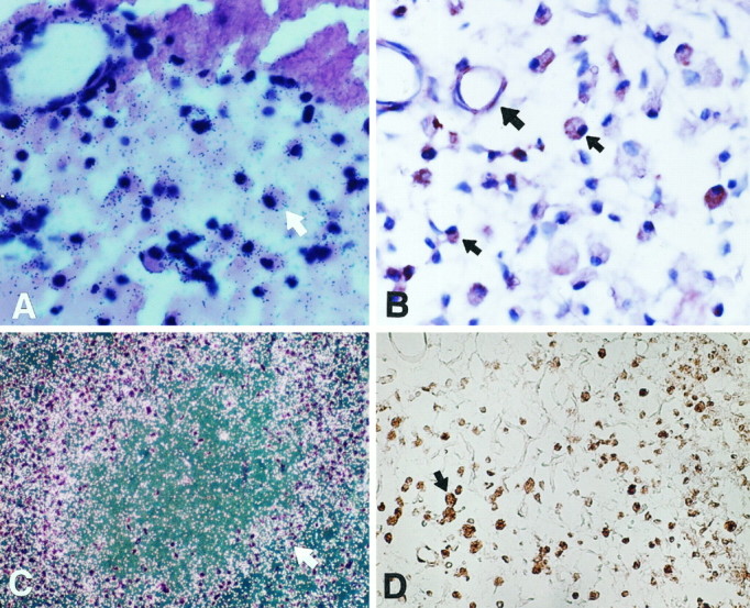
Pleiotrophin (PTN) is a heparin-binding, 18 kDa secretory protein that functions to induce mitogenesis, angiogenesis, differentiation, and transformation in vitro. PTN gene (Ptn) expression is highly regulated during development and is highest at sites in which mitogenesis, angiogenesis, and differentiation are active. In striking contrast, with the exception of the neuron, the Ptn gene is only minimally expressed in adults. We now demonstrate that Ptn gene expression is strikingly upregulated within 3 d in OX42-positive macrophages, astrocytes, and endothelial cells in areas of developing neovasculature after focal cerebral ischemia in adult rat. Ptn gene expression remains upregulated in these same cells and sites 7 and 14 d after ischemic injury. However, expression of the Ptn gene is significantly decreased in cortical neurons 6 and 24 hr after injury and is undetectable in degenerating neurons at day 3. Neurons in contralateral cortex continue to express Ptn in levels equal to control, uninjured brain. It is suggested that PTN may have a vital role in neovascular formation in postischemic brain and that postischemic brain is an important model in which to analyze sequential gene expression in developing neovasculature. In contrast, Ptn gene expression in injured neurons destined not to recover is strikingly reduced, and potentially its absence may contribute to the failure of the neuron to survive.


Figures

Fig. 1.
Fig. 1.
Top. Sections of a brain from a sham-operated rat hybridized with 35S-Ptnantisense cRNA probe (A, dark field; B, bright field) or 35S-Ptn sense cRNA probe (C, bright field). Ptn hybridization signal was highly expressed in neurons of cortex (large arrow). A little signal was found in glial cells (small arrow) and microvascular endothelium (medium arrow). D (bright field), Section hybridized with 35S-PTN antisense cRNA shows that PTN mRNA decreases to a greater degree in degenerating neurons in the ischemic core (C) then in the periphery (P) of the infarct compared with normal neurons (N) 6 hr after reperfusions. Magnification:A–C, 200×; D, 100×.
Fig. 3.
Fig. 3.
Sections of a brain 3 d after ischemia hybridized with 35S-Ptn antisense cRNA (A, B) and immunostaining with anti-PTN antibody (C) or anti-GFAP antibody (D). Ptn hybridization signal was predominantly expressed in the neurons (large arrow) and glial cells (small arrow) along the border of the infarct and blood vessels (arrowhead) immediately adjacent to the infarct (top right in A, dark field, B, bright field). PTN immunoreactivity was detected in glial cells (large arrow) at the border of the infarct (C), which corresponded to the hypertrophic anti-GFAP(+) astrocytes in the similar area on day 3 (D, large arrow); the small arrow denotes neurons under degeneration. Magnification, 200×.
Fig. 4.
Fig. 4.
Sections of an ischemic brain on day 3 hybridized with 35S-Ptn antisense cRNA (A, C, dark field; B, D, bright field). PTN transcripts were found in the endothelium of blood vessels (large arrow) and glial cells (small arrow). Endothelial sprouts were seen in Aand B (large arrow). Magnification, 200×.
Fig. 5.
Fig. 5.
Sections of an ischemic brain on day 3. A, B, PTN immunoreactivity was found in endothelial cells (small arrow) and macrophages (large arrow) in the infarcted region after staining with anti-PTN antibody. C, D, Macrophages surrounding blood vessel (large arrow) were identified by immunostaining with anti-OX42 antibody (C, arrow, frozen section) and histochemistry staining with GSA-IB4(D, arrow). Magnification: A, B, 400×;C, D, 200×.
Fig. 6.
Fig. 6.
Sections of ischemic brain on days 3, 7, and 14.A, 35S-Ptn cRNA hybridization signals were detected in macrophages (arrow) near the infarcted area (top) on day 3 (bright field).B, A number of macrophages with PTN immunoreactivity (small arrow) with variable morphology in the infarcted area on day 7 (large arrow denotes a blood vessel).C, 35S-Ptn antisense cRNA hybridization signals were detected in numerous macrophages (arrow) surrounding residual necrotic tissue at the infarcted region on day 14 (bright field). D, A number of macrophages stained with GSA-IB4 with various morphological features in the infarct on day 14. Magnification:A, B, 400×; C, D, 100×.

Similar articles

Cited by

References

    1. Chauhan AK, Li YS, Deuel TF. Pleiotrophin transformations NIH 3T3 cells and induced tumors in nude mice. Proc Natl Acad Sci USA. 1993;90:679–682. - PMC - PubMed
    1. Choudhuri R, Zhang HT, Donnini S, Ziche M, Bicknell R. An angiogenic role for the neurokines midkine and pleiotrophin in tumorigenesis. Cancer Res. 1997;57:1814–1819. - PubMed
    1. Courty J, Dauchel MC, Caruelle D, Perderiset M, Barritault D. Mitogenic properties of a new endothelial cell growth factor related to pleiotrophin. Biochem Biophys Res Commun. 1991;180:145–151. - PubMed
    1. Czubayko F, Schulte AM, Berchem GJ, Wellstein A. Melanoma angiogenesis and metastasis modulated by ribozyme targeting of the secreted growth factor pleiotrophin. Proc Natl Acad Sci USA. 1996;93:14753–14758. - PMC - PubMed
    1. Fang W, Hartmann N, Chow DT, Riegel AT, Wellstein A. Pleiotrophin stimulates fibroblasts and endothelial and epithelial cells and is expressed in human cancer. J Biol Chem. 1992;267:25889–25897. - PubMed

Publication types

LinkOut - more resources