. 1998 May 15;18(10):3803-15.
doi: 10.1523/JNEUROSCI.18-10-03803.1998.

Long-distance axonal regeneration in the transected adult rat spinal cord is promoted by olfactory ensheathing glia transplants

Affiliations

Long-distance axonal regeneration in the transected adult rat spinal cord is promoted by olfactory ensheathing glia transplants

A Ramón-Cueto et al. J Neurosci. .

Abstract

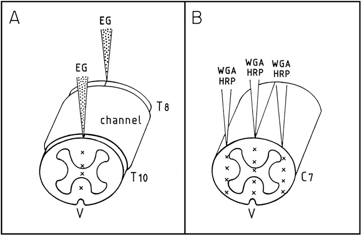
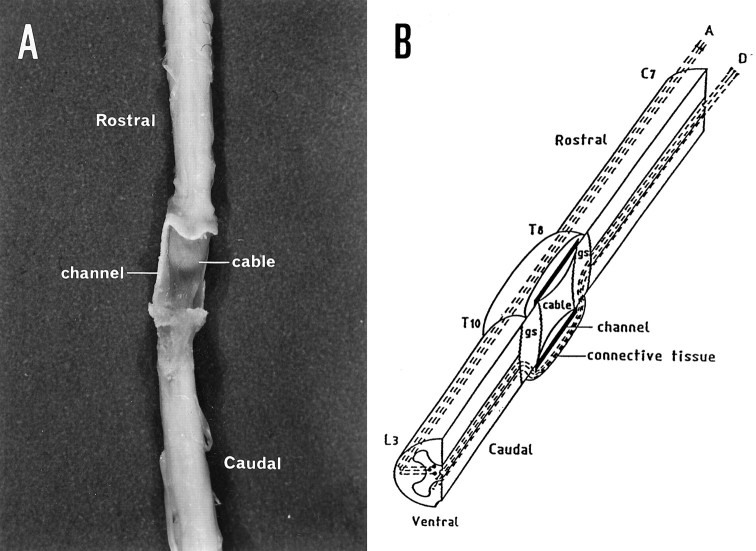
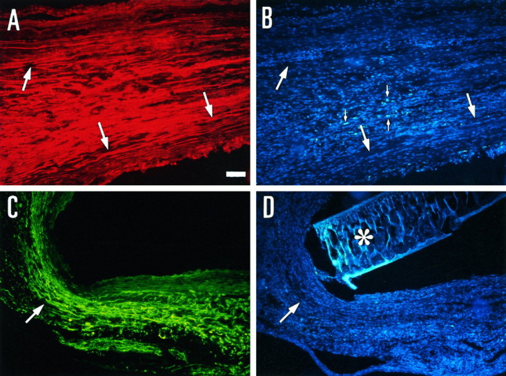
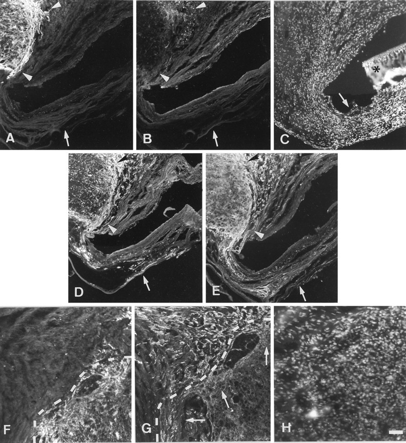
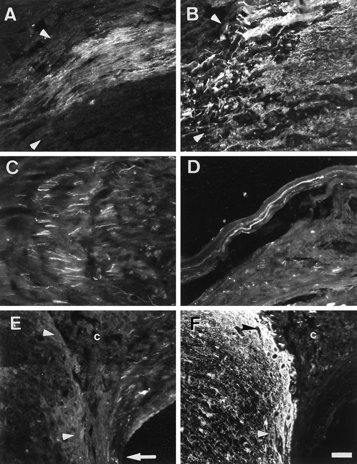
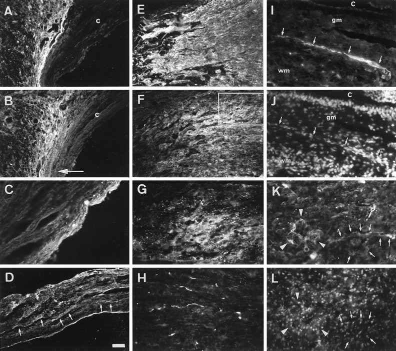
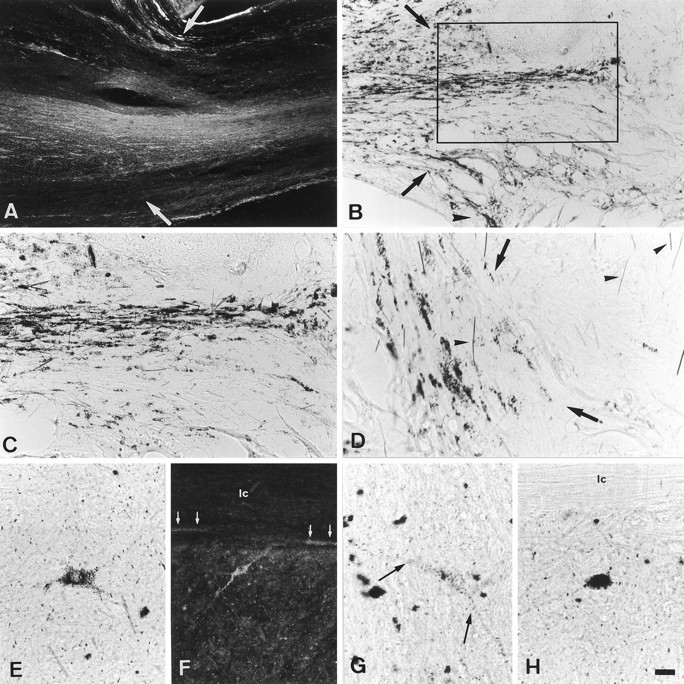
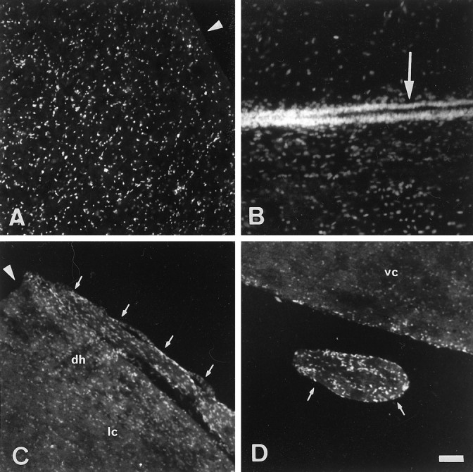
The lack of axonal regeneration in the injured adult mammalian spinal cord leads to permanent functional impairment. To induce axonal regeneration in the transected adult rat spinal cord, we have used the axonal growth-promoting properties of adult olfactory bulb ensheathing glia (EG). Schwann cell (SC)-filled guidance channels were grafted to bridge both cord stumps, and suspensions of pure (98%) Hoechst-labeled EG were stereotaxically injected into the midline of both stumps, 1 mm from the edges of the channel. In EG-transplanted animals, numerous neurofilament-, GAP-43-, anti-calcitonin gene-related peptide (CGRP)-, and serotonin-immunoreactive fibers traversed the glial scars formed at both cord-graft interfaces. Supraspinal serotonergic axons crossed the transection gap through connective tissue bridges formed on the exterior of the channels, avoiding the channel interior. Strikingly, after crossing the distal glial scar, these fibers elongated in white and periaqueductal gray matter, reaching the farthest distance analyzed (1.5 cm). Tracer-labeled axons present in SC grafts were found to extend across the distal interface and up to 800 microm beyond in the distal cord. Long-distance regeneration (at least 2.5 cm) of injured ascending propriospinal axons was observed in the rostral spinal cord. Transplanted EG migrated longitudinally and laterally from the injection sites, reaching the farthest distance analyzed (1.5 cm). They moved through white matter tracts, gray matter, and glial scars, overcoming the inhibitory nature of the CNS environment, and invaded SC and connective tissue bridges and the dorsal and ventral roots adjacent to the transection site. Transplanted EG and regenerating axons were found in the same locations. Because EG seem to provide injured spinal axons with appropriate factors for long-distance elongation, these cells offer new possibilities for treatment of CNS conditions that require axonal regeneration.


Figures

Fig. 1.
Fig. 1.
A, Diagram showing the four sites (x) in the midline of both spinal cord stumps where suspensions of ensheathing glia (EG) were grafted.B, Diagram illustrating the 13 spinal cord sites (x) of cervical segment 7 (C7) where wheat germ agglutinin–horseradish peroxidase (WGA–HRP) was injected 6 weeks after EG transplantation. T8 and T10, Thoracic segments 8 and 10; V, ventral.
Fig. 2.
Fig. 2.
A, Dorsal surface of a perfused EG-transplanted spinal cord 6 weeks after surgery. To expose the SC cable, we removed the dorsal part of the channel. The cable was firmly attached to both rostral and caudal spinal cord stumps.B, Diagram showing a sagittal hemisection of a grafted spinal cord 6 weeks after transplantation. Two bridges were present between rostral and caudal cord stumps: (1) a SC- and EG-containing cable inside the channel and (2) a connective tissue layer containing EG around the channel. Dashed lines represent regenerating ascending (A) and some of the descending (D) fibers. Because of space constraints, the ascending fibers and some of the descending fibers were not drawn through the SC bridge. C7, Cervical segment 7; gs, glial scar; L3, lumbar segment 3; T8 and T10, thoracic segments 8 and 10.
Fig. 3.
Fig. 3.
Regenerating axons growing inside either an SC- and EG-containing cable (A, B) or an EG-containing connective tissue bridge (C,D). A, Regenerating axons visualized by immunolabeling with anti-GAP-43 antibody. B, Hoechst-labeled EG observed in the same field shown inA. C, Regenerating axons immunolabeled with anti-neurofilament antibody. D, Hoechst-labeled EG present in the same field shown in C. Large arrows point to EG (B, D) aligned along bundles of regenerating axons (A,C). Small arrows in Bpoint to Hoechst-labeled nuclei of macrophages and microglial cells. Note the difference in shape and color of these cells compared with that of EG. A portion of the channel, which autofluoresces with the filter used, was retained in D(asterisk). Scale bar, 60 μm.
Fig. 4.
Fig. 4.
Regenerating axons crossing the gliotic tissue created at both graft–cord interfaces after EG transplantation.AE, Consecutive spinal cord sagittal sections showing the interfaces between the rostral cord stump and either SC and EG cables or EG connective tissue bridges. Sections were immunolabeled for GFAP (A), ED1 (B), neurofilament (D), and GAP-43 (E). EG were visualized by Hoechst labeling (C). A portion of the channel, which autofluoresces with the filter used, was retained in C(asterisk). Arrowheads inA, B, D, andE point to the cord–cable interface;arrows in AE mark the EG-containing connective tissue bridge.FH, Consecutive spinal cord sections showing the caudal cord–cable interface. Dashed lines(F, G) indicate the interface. Sections were immunolabeled for GFAP (F) and GAP-43 (G). Hoechst-labeled EG (H) are in the same region shown inF. Arrows in G point to GAP-43-labeled fibers crossing the cord–cable interface and in the gliotic tissue. Note the presence of EG in the gliotic tissue of both interfaces (C, H) and in both SC-containing cables and connective tissue bridges (C). Scale bars:AE, 125 μm;FH, 90 μm.
Fig. 5.
Fig. 5.
Regeneration of CGRP-positive fibers in the transected spinal cord after EG transplantation. A,B, Consecutive spinal cord sections showing CGRP-immunoreactive fibers (A) crossing the GFAP-positive gliotic tissue (B) at the caudal cable–cord interface (arrowheads) from the dorsal columns. C, D, CGRP-positive fibers regenerating through an SC and EG cable (C) or a bridge of EG connective tissue (D).E, F, Consecutive sections immunolabeled for CGRP (E) or GFAP (F).E shows CGRP-immunoreactive axons growing inside an SC- and EG-containing cable (c) and a connective tissue bridge (arrow), near the glial scar of the rostral interface (arrowheads in E andF). Scale bars: A,B, 85 μm; C, D, 50 μm;E, F, 90 μm.
Fig. 6.
Fig. 6.
Regeneration of serotonergic axons after EG transplantation. A, B, Consecutive spinal cord sections immunolabeled for GFAP (A) and serotonin (B). Serotonergic fibers grow through the GFAP-positive gliotic tissue at the rostral transection site and reach the rostral cord–cable. Most fibers grow ventrally toward the connective tissue that surrounds the channel (arrow).c, Cable. C, Serotonergic axons at the periphery of an SC and EG cable 1 mm from the cord–cable interface.D, Serotonergic axons (arrows) regenerating through the EG-containing connective tissue surrounding the channel. E, F, Consecutive spinal cord sections immunolabeled with anti-GFAP (E) and anti-serotonin (F) showing serotonergic fibers in the glial scar of the caudal stump. G, Higher magnification of the serotonergic fibers in the boxed region in F. H, Serotonergic axons regenerating through the ventral columns at the L2 level.I, Section of the distal spinal cord at the level of L2 immunolabeled for serotonin. J, Hoechst-labeled EG in the same field shown in I. Arrows(I, J) point to one serotonin-immunostained axonal bundle at the periaqueductal gray matter (gm) bordering the white matter (wm) of the ventral columns. Note the close association of the fibers and EG (compare arrows inI, J). c, Central canal. K, Serotonin-immunoreactive fibers in the ventral horn of the caudal cord stump. Note that some immunopositive fibers delineate the dendrites and bodies of neurons; arrowspoint to a neuron the dendrites and body of which are outlined by serotonergic immunoreactivity, and arrowheads point to serotonin immunostaining surrounding a group of neurons.L. Same field shown in K revealing Hoechst-labeled EG. The Hoechst-labeled nuclei colocalize with the serotonergic fibers associated with the neurons shown inK (arrows and arrowheads). Scale bars: A, B, E,F, I, J, 100 μm;C, 50 μm; D, 90 μm; G,H, K, L, 45 μm.
Fig. 7.
Fig. 7.
Photomicrographs of WGA–HRP tracing visualized by TMB histochemistry in experimental and control animals. Spinal cords were cut in the horizontal direction. A, Dark-field photograph showing labeled axons entering an SC- and EG-containing cable. Arrows point to the rostral cord (left)–cable (right) interface.B, Bright-field photograph illustrating labeled axons entering the caudal spinal cord stump. Arrows indicate the cable (left)–host (right) interface. The arrowhead points to labeled axons growing through the connective tissue bridge surrounding the channel. C, Higher magnification of boxed area in Bshowing the regenerating axons entering the caudal cord stump.D, In control animals, labeled axons are seen in the distal-most region of the cable but not in the distal cord stump. The cable (left)–cord (right) interface is marked by arrows. Arrowheads indicate nonspecific TMB crystals. EH, Photomicrographs of neurons retrogradely traced with WGA–HRP in the caudal cord stump. F is a dark-field andE, G, and H are bright-field photomicrographs. Labeled neurons are in lamina V of T11 (E) and L1 (F) cord segments. Arrows in F point to labeled axons running in the lateral columns (lc). Labeled neurons are in lamina VII of L1 (G) and L3 (H) cord segments. The processes (arrows) but little of the cell body of the neuron are present in the section shown in G. In H, the labeled neuron is located near the lateral columns (lc). Scale bars: A, B, 160 μm; C, F, 80 μm;D, 50 μm; E, G,H, 40 μm.
Fig. 8.
Fig. 8.
EG migration through the transected spinal cord visualized by Hoechst nuclear labeling. A, Labeled nuclei in the lateral columns of the caudal cord stump. Thearrowhead points to a distance of 1.5 cm caudal to the edge of the guidance channel, the farthest distance analyzed.B, Labeled EG nuclei in the ependymal layer (arrow) and the periaqueductal gray matter of the caudal cord stump. C, D, Labeled EG nuclei in a dorsal root (arrows in C) and a ventral root (arrows in D). dh, Dorsal horn; lc, lateral column; vc, ventral column. The arrowhead in C points to a distance of 1.5 cm rostral to the edge of the channel, the farthest distance analyzed. Notice that some regions lack Hoechst-labeled nuclei (see top half inB, lateral columns in C, and ventral columns and root in D). Scale bars: A,C, D, 100 μm; B, 50 μm.

Similar articles

Cited by

References

    1. Baird A, Klagsbrun M. The fibroblast growth factor family. Ann NY Acad Sci. 1991;638:239–243. - PubMed
    1. Bandtlow C, Zachleder T, Schwab ME. Oligodendrocytes arrest neurite growth by contact inhibition. J Neurosci. 1990;10:3837–3848. - PMC - PubMed
    1. Baron-Van Evercooren A, Gansmuller A, Clerin E, Gumpel M. Hoechst 33342 a suitable fluorescent marker for Schwann cells after transplantation in the mouse spinal cord. Neurosci Lett. 1991;131:241–244. - PubMed
    1. Bixby JL, Lilien J, Reichardt LF. Identification of the major proteins that promote neuronal process outgrowth on Schwann cells in vitro. J Cell Biol. 1988;107:353–361. - PMC - PubMed
    1. Bovolenta P, Wandosell F, Nieto-Sampedro M. CNS glial scar tissue: a source of molecules which inhibit central neurite outgrowth. Prog Brain Res. 1992;94:367–379. - PubMed

Publication types

MeSH terms

Substances