. 1998 May;9(5):977-91.
doi: 10.1091/mbc.9.5.977.

Saccharomyces cerevisiae cells with defective spindle pole body outer plaques accomplish nuclear migration via half-bridge-organized microtubules

Affiliations
Free PMC article

Saccharomyces cerevisiae cells with defective spindle pole body outer plaques accomplish nuclear migration via half-bridge-organized microtubules

A Brachat et al. Mol Biol Cell. 1998 May.
Free PMC article

Abstract

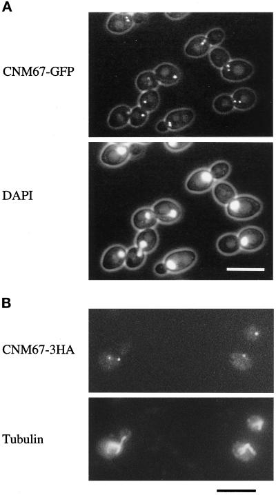
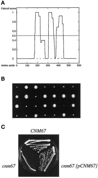
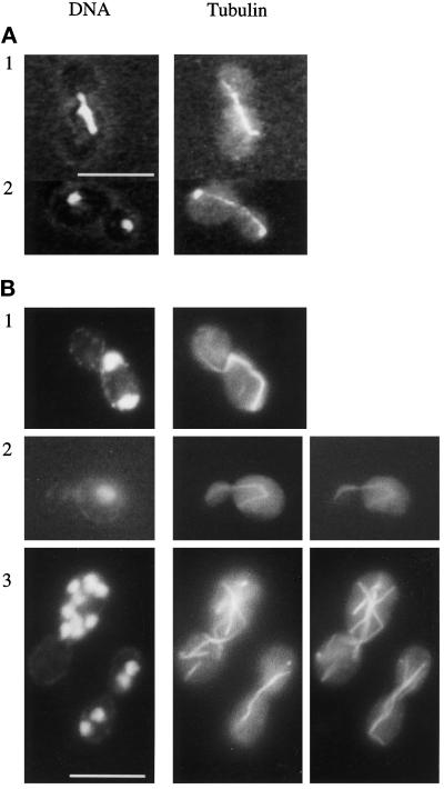
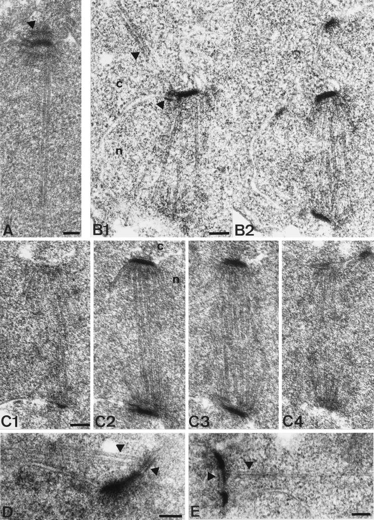
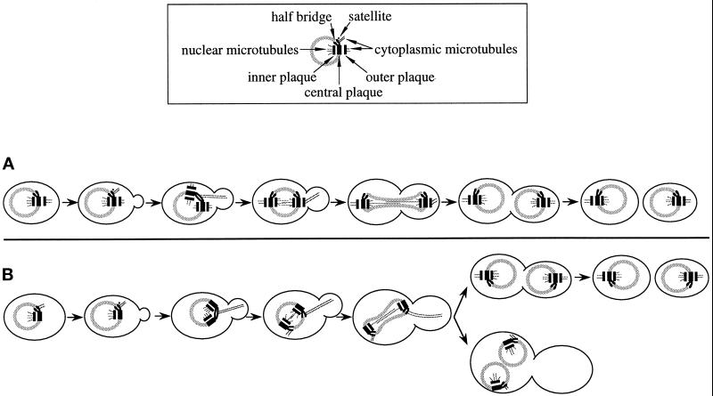
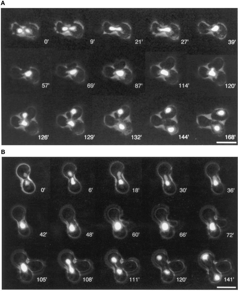
Cnm67p, a novel yeast protein, localizes to the microtubule organizing center, the spindle pole body (SPB). Deletion of CNM67 (YNL225c) frequently results in spindle misorientation and impaired nuclear migration, leading to the generation of bi- and multinucleated cells (40%). Electron microscopy indicated that CNM67 is required for proper formation of the SPB outer plaque, a structure that nucleates cytoplasmic (astral) microtubules. Interestingly, cytoplasmic microtubules that are essential for spindle orientation and nuclear migration are still present in cnm67Delta1 cells that lack a detectable outer plaque. These microtubules are attached to the SPB half- bridge throughout the cell cycle. This interaction presumably allows for low-efficiency nuclear migration and thus provides a rescue mechanism in the absence of a functional outer plaque. Although CNM67 is not strictly required for mitosis, it is essential for sporulation. Time-lapse microscopy of cnm67Delta1 cells with green fluorescent protein (GFP)-labeled nuclei indicated that CNM67 is dispensable for nuclear migration (congression) and nuclear fusion during conjugation. This is in agreement with previous data, indicating that cytoplasmic microtubules are organized by the half-bridge during mating.


Figures

Figure 1
Figure 1
Subcellular localization of functional Cnm67p fusion proteins visualized by fluorescence microscopy. (A) Cnm67-GFPp fluorescence is visible at one or two spots at the nuclear periphery, consistent with a localization to the SPB. Nuclear DNA was stained with DAPI. (B) Immunofluorescence microscopy of Cnm67–3HAp–labeled cells. Dot-shaped staining with anti-HA antibody coincides with the spindle poles visualized by tubulin staining with anti-tubulin antibody.
Figure 2
Figure 2
Cnm67p coiled-coil prediction, tetrad analysis, and complementation test. (A) Probability plot for coiled-coil formation of the 581 amino-acid sequence of Cnm67p. The central part of the protein was predicted to contain three α helical coiled-coil regions (residues 174–226, p = 1; residues 306–357, p = 1; residues 382–448, p = 0.924) by the Paircoil program (Berger et al., 1995). The CNM67 sequence is available from EMBL/GenBank/DDBJ under accession number Z71501. (B) Tetrad dissection of a heterozygous CNM67-deleted strain (ABY101). Tetrad colonies were transferred on G418 containing medium to test for segregation of the kanMX4 gene deletion marker. Slow growing colonies always carried the cnm67::kanMX4 allele (cnm67Δ1). (C) Complementation of the cnm67Δ1 growth defect by transformation with a plasmid carrying the CNM67 wild-type allele.
Figure 3
Figure 3
Nuclear distribution and microtubule structure in wild-type and cnm67Δ1. Cells were double stained for nuclear DNA with DAPI and for tubulin with anti-tubulin antibody. (A) Wild-type. (B) cnm67Δ1 cells. Panels B2 and B3 show different focal planes of the respective cells to visualize the complete microtubular structures. B1 and B2 show cytoplasmic microtubules that direct from one spindle pole into the bud. The spindle in B1 is highly elongated although it is restricted to the mother cell. Panel B3 depicts multinucleated cells. Bars, 10 μm.
Figure 4
Figure 4
Thin section electron microscopy of wild-type and cnm67Δ1 spindles. The nucleus is marked by ’n’ and the cytoplasm by ’c’. (A) SPB of a wild-type cell with elongated spindle. The SPB half-bridge as well as the central, inner, and outer plaque of the SPB are visible. The arrowhead shows the outer plaque. Cytoplasmic microtubules do not lie within the plane of the section. (B1–2) Serial thin sections of a complete cnm67Δ1 spindle after SPB separation. Unlike in wild-type cells, cytoplasmic microtubules are attached to the half-bridge of the upper SPB. Arrowheads show cytoplasmic microtubules and the half-bridge. (C1–2) Serial thin sections of a cnm67Δ1 spindle. The SPB outer plaque is not detectable in any of the sections, although the half-bridge, central, and inner plaques appear normal. The same was found for all cnm67Δ1 SPBs that we investigated (n>300). (D) Cytoplasmic microtubules are attached to the half-bridge of a cnm67Δ1 cell with a single SPB, presumably a G1 cell. Arrowheads show the half-bridge and cytoplasmic microtubules. (E) Side- by-side SPBs are connected by a bridge that nucleates cytoplasmic microtubules directing into the bud. The bridge and cytoplasmic microtubules are indicated. Bars, 0.1 μm.
Figure 5
Figure 5
Model for a salvage mechanism for spindle orientation and nuclear migration in cnm67Δ1 cells. (A) Wild-type: SPB half-bridges and bridges bind cytoplasmic microtubules only when the satellite forms and SPB duplication takes place. After SPB separation, cytoplasmic microtubules are no longer found at the half-bridge (Byers and Goetsch, 1975). During all cell cycle stages, the outer plaque acts as an attachment site. The first phase of nuclear migration begins in S when cytoplasmic microtubules originate from the bridge of side-by-side SPBs. The second phase of nuclear migration in G2/M coincides with cytoplasmic microtubule attachment at the outer plaque. (B) In cnm67Δ1 cells cytoplasmic microtubules originate from the half-bridge throughout the cell cycle while the outer plaque is much reduced or absent and thus incapable of microtubule binding. Consequently, the second phase of nuclear migration is impaired, frequently leading to nuclear division within the mother cell. However, half-bridge–organized cytoplasmic microtubules still have a limited capability to mediate spindle positioning and nuclear migration forces that allow many cells to finally migrate the daughter nucleus into the bud. Cells in which nuclear migration failed double in ploidy and often undergo another round of nuclear division.
Figure 6
Figure 6
Time-lapse fluorescence video microscopy of wild-type and cnm67Δ1 conjugations. Nuclei of haploid cells were labeled with a strongly fluorescent histone H4-GFPp (Hhf2-GFPp). (A) A nuclear fusion event of wild-type cells is followed by the first mitotic division of the zygote nucleus. Both daughter nuclei are correctly segregated between the zygote and the bud. Nuclear division is then followed by cytokinesis. (B) Conjugation of cnm67Δ1 cells. Nuclear migration (congression) before nuclear fusion and nuclear fusion occur as in wild-type cells in the central region of the zygote and also with about the same dynamics. The subsequent mitotic division, however, fails to segregate one of the daughter nuclei into the zygote bud. Presumably, congression is not affected because it is mediated by half-bridge–organized microtubules as in wild-type, whereas the first mitotic nuclear migration is impaired by the outer plaque defect of cnm67Δ1 cells. Cells that surrounded mating cells in panels A and B were removed electronically for clarity. Bars, 10 μm.

References

    1. Adams AE, Pringle JR. Relationship of actin and tubulin distribution to bud growth in wild-type and morphogenetic-mutant Saccharomyces cerevisiae. J Cell Biol. 1984;98:934–945. - PMC - PubMed
    1. Berger B, Wilson DB, Wolf E, Tonchev T, Milla M, Kim PS. Predicting coiled coils by use of pairwise residue correlations. Proc Natl Acad Sci USA. 1995;92:8259–8263. - PMC - PubMed
    1. Baudin A, Ozier KO, Denouel A, Lacroute F, Cullin C. A simple and efficient method for direct gene deletion in Saccharomyces cerevisiae. Nucleic Acids Res. 1993;21:3329–3330. - PMC - PubMed
    1. Brinkley BR. Microtubule organizing centers. Annu Rev Cell Biol. 1985;1:145–172. - PubMed
    1. Bullitt E, Rout MP, Kilmartin JV, Akey CW. The yeast spindle pole body is assembled around a central crystal of Spc42p. Cell. 1997;89:1077–1086. - PubMed

Publication types

MeSH terms