. 1998 May;9(5):1195-207.
doi: 10.1091/mbc.9.5.1195.

A role for katanin-mediated axonemal severing during Chlamydomonas deflagellation

Affiliations
Free PMC article

A role for katanin-mediated axonemal severing during Chlamydomonas deflagellation

T A Lohret et al. Mol Biol Cell. 1998 May.
Free PMC article

Abstract

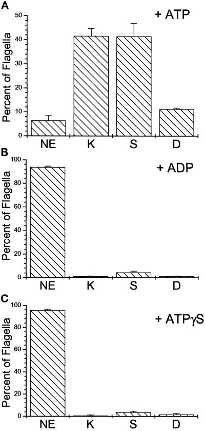
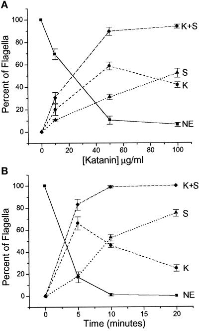
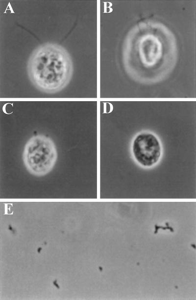
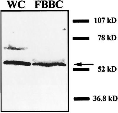
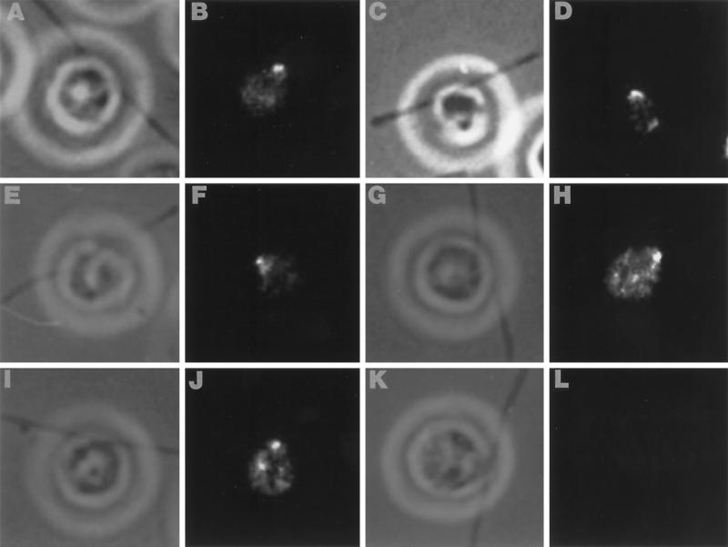
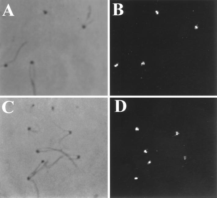
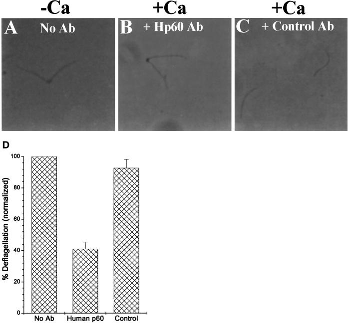
Deflagellation of Chlamydomonas reinhardtii, and other flagellated and ciliated cells, is a highly specific process that involves signal-induced severing of the outer doublet microtubules at a precise site in the transition region between the axoneme and basal body. Although the machinery of deflagellation is activated by Ca2+, the mechanism of microtubule severing is unknown. Severing of singlet microtubules has been observed in vitro to be catalyzed by katanin, a heterodimeric adenosine triphosphatase that can remove tubulin subunits from the walls of stable microtubules. We found that purified katanin induced an ATP-dependent severing of the Chlamydomonas axoneme. Using Western blot analysis and indirect immunofluorescence, we demonstrate that Chlamydomonas expresses a protein that is recognized by an anti-human katanin antibody and that this protein is localized, at least in part, to the basal body complex. Using an in vitro severing assay, we show that the protein(s) responsible for Ca2+-activated outer doublet severing purify with the flagellar-basal body complex. Furthermore, deflagellation of purified flagellar-basal body complexes is significantly blocked by the anti-katanin antibody. Taken together, these data suggest that a katanin-like mechanism may mediate the severing of the outer doublet microtubules during Chlamydomonas deflagellation.


Figures

Figure 1
Figure 1
The machinery of microtubule-severing isolates with the FBBC. Representative phase-contrast micrographs of purified FBBCs immediately after purification (A) and after calcium treatment (B). Each flagella is connected to a basal body and the two basal bodies are linked by connecting fibers. FBBCs were purified on a Percoll gradient after detergent lysis of GLE-treated wild-type cells and resuspended in calcium-free DB (see MATERIALS AND METHODS). Deflagellation of the FBBCs was induced by addition of CaCl2 to a final free Ca2+ concentration of 1 μM. As shown in panel B, the addition of micromolar free Ca2+ resulted in the complete severing of the axonemes from the basal bodies. The flagella and basal bodies are labeled F and B, respectively.
Figure 2
Figure 2
Katanin severs Chlamydomonas axonemal microtubules. NP-40–permeabilized cells (≈1 × 10⁵ cells/ml) were incubated with purified katanin (100 μg/ml) plus 2.5 mM ADP (A) or 2.5 mM ATP (B–D) for 10 min. In the presence of ATP, katanin causes kinking (B), severing (C), and complete deflagellation (D) of individual flagella. (E and F) Katanin induces kinking and breakage of isolated flagellar axonemes. Flagella were excised from permeabilized wild-type cells by 1 × 10⁻⁶ M free Ca2+ and treated with either 5 mM ATP (E) or 5 mM ATP plus katanin (F) (100 μg/ml). As shown, the axonemes are fragmented only in the presence of both katanin and ATP.
Figure 3
Figure 3
Katanin-mediated flagellar microtubule severing is supported by ATP but not by either ADP or ATPγS. The percentage of flagella scored in each of the categories illustrated in Figure 3 was determined in the presence of katanin (100 μg/ml) plus the indicated nucleotide (2.5 mM). As shown, katanin-induced flagellar axonemal microtubule kinking and breakage was supported by ATP (A) but not by ADP or ATPγS (B and C). Bars represent the average ± SE, n = 4; 50 cells counted for each experiment. All other conditions as in Figure 2.
Figure 4
Figure 4
Katanin-mediated breakage of Chlamydomonas flagella is both dose and time dependent. (A) NP-40–permeabilized wild-type cells (≈1 × 10⁵ cells/ml) were incubated with purified katanin plus ATP for 10 min at various katanin concentrations (μg/ml). The percent of kinked and/or severed flagella (scored and quantified as in Figure 3) after 10 min of incubation was determined at various katanin concentrations (μg/ml). Note that flagella scored as deflagellated were added to those scored as severed. (B) Wild-type cells were permeabilized with 0.05% NP-40, 5 mM ATP, 100 μg/ml katanin for various amounts of time. At the indicated time points, katanin activity was stopped by addition of 0.8% glutaraldehyde and the flagella were assayed as in panel A. For assays at 20 min, 5 mM ATP was added after 10 min of incubation. For parts A and B, data points represent the average ± SE, n = 3–5; minimum of 50 cells counted for each experiment.
Figure 5
Figure 5
Katanin induces kinking and severing of fa1–1 flagella. NP-40 (0.05%)-permeabilized fa1 mutant cells (≈1 × 10⁵ cells/ml) were incubated with purified katanin (100 μg/ml) plus 5 mM ADP (A) or 5 mM ATP (B–D) for 10 min. In the presence of ATP, katanin causes kinking (B), severing (C), and deflagellation (D) of fa1–1 flagella. (E) Flagellar preparations made from the katanin-treated fa1 cells consisted of small, kinked flagellar fragments resembling isolated wild-type flagella treated with katanin (Figure 2F).
Figure 6
Figure 6
Anti-human p60 antibodies strongly recognize a ≈55-kDa Chlamydomonas protein in both whole-cell and flagellar–basal body protein preparations. Whole-cell and FBBC proteins (10 μg) were separated on a 4–15% SDS-PAGE gel and analyzed by Western blot with an affinity-purified antibody to the 60-kDa subunit of human katanin. Detection was with an alkaline phosphatase-conjugated secondary antibody.
Figure 7
Figure 7
Indirect immunofluorescence of whole cells demonstrates immunoreactivity near the basal bodies, but not along the length of the axoneme. Alternating phase contrast (A, C, E, G, and I) and anti-human p60 katanin fluorescence images (B, D, F, H, and J) of several different cells illustrating strong immunoreactivity at the base of the FBBC. Immune detection is with a FITC-conjugated secondary antibody. (K and L) Representative phase contrast and fluorecence images of a control cell that was treated with secondary antibody.
Figure 8
Figure 8
Anti-human p60 antibodies specifically recognize basal body-localized epitopes in preparations of purified FBBCs. Phase contrast (A and C) and anti-human p60 katanin fluorescence images (B and D) of FBBCs illustrating intense staining of the basal bodies. No staining was observed along the length of the axonemes. Controls were performed with secondary antibody alone and showed no fluorescent staining of the basal bodies.
Figure 9
Figure 9
Anti-human p60 antibodies inhibit deflagellation of isolated FBBCs. Purified FBBCs were treated with either no antibody (A), 60 μg/ml anti-human p60 antibody (B), or an equivalent amount of control antibodies (C) for 15 min and then assayed for Ca-induced deflagellation by addition of 1 μM free Ca2+. (D) The percent deflagellation of each sample was calculated as described (see MATERIALS AND METHODS) and normalized to the untreated sample.

References

    1. Blum JJ. Existence of a breaking point in cilia and flagella. J Theor Biol. 1971;33:257–263. - PubMed
    1. Dutcher SK. Purification of basal bodies and basal body complexes from Chlamydomonas reinhardtii. In: Dentler W, Witman G, editors. Cilia and Flagella. San Diego, CA: Academic Press; 1995. . Methods in Cell Biology 47, 323–334. - PubMed
    1. Dye RB, Flicker PF, Lien DY, Williams RC. End-stabilized microtubules observed in vitro: stability, subunit interchange, and breakage. Cell Motil Cytoskeleton. 1992;21:171–186. - PubMed
    1. Fabiato. A. Computer programs for calculating total from free or free from specified total ionic concentrations in aqueous solution containing multiple metals and ligands. Methods Enzymol. 1988;157:378–417. - PubMed
    1. Fechter J, Schoneberg A, Schatten G. Excision and disassembly of sperm tail microtubules during sea urchin fertilization: Requirements for microtubule dynamics. Cell Motil Cytoskeleton. 1996;35:281–288. - PubMed

Publication types

LinkOut - more resources