. 1998 May;64(5):1766-72.
doi: 10.1128/AEM.64.5.1766-1772.1998.

Molecular analysis of a laccase gene from the white rot fungus Pycnoporus cinnabarinus

Affiliations

Molecular analysis of a laccase gene from the white rot fungus Pycnoporus cinnabarinus

C Eggert et al. Appl Environ Microbiol. 1998 May.

Abstract

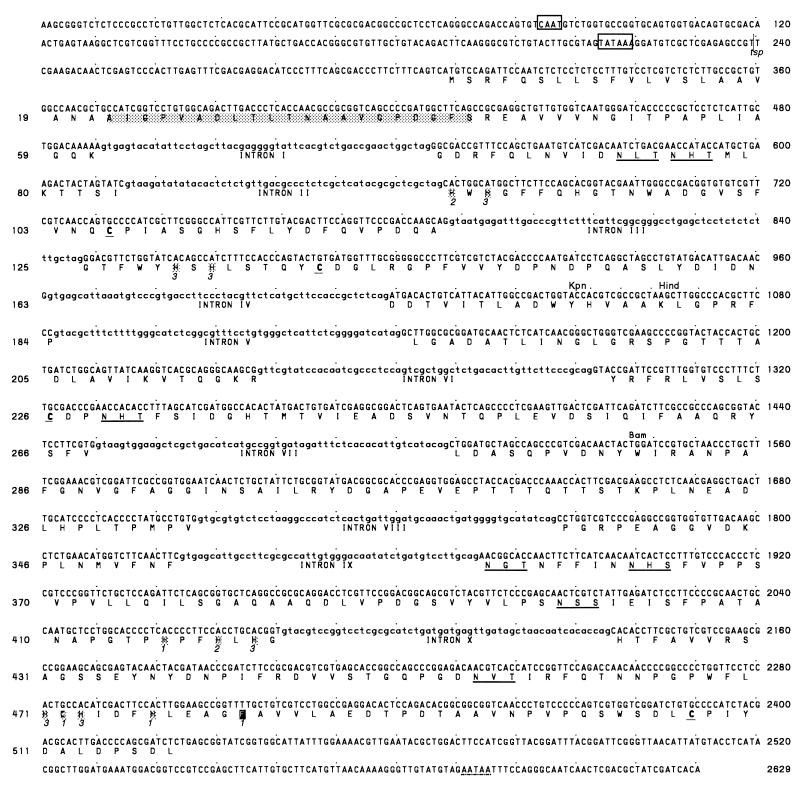
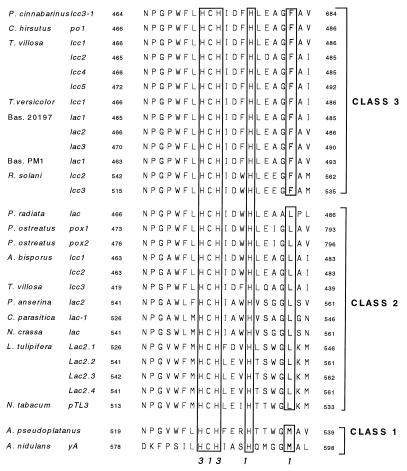
It was recently shown that the white rot basidiomycete Pycnoporus cinnabarinus secretes an unusual set of phenoloxidases when it is grown under conditions that stimulate ligninolysis (C. Eggert, U. Temp, and K.-E. L. Eriksson, Appl. Environ. Microbiol. 62:1151-1158, 1996). In this report we describe the results of a cloning and structural analysis of the laccase-encoding gene (lcc3-1) expressed by P. cinnabarinus during growth under xylidine-induced conditions. The coding region of the genomic laccase sequence, which is preceded by the eukaryotic promoter elements TATA and CAATA, spans more than 2,390 bp. The corresponding laccase cDNA was identical to the genomic sequence except for 10 introns that were 50 to 60 bp long. A sequence analysis indicated that the P. cinnabarinus lcc3-1 product has a Phe residue at a position likely to influence the reduction-oxidation potential of the enzyme's type 1 copper center. The P. cinnabarinus lcc3-1 sequence was most similar to the sequence encoding a laccase from Coriolus hirsutus (level of similarity, 84%).


Figures

FIG. 1
FIG. 1
Strategy used for PCR cloning of the laccase-encoding cDNA from P. cinnabarinus and oligonucleotide primer sequences. The boxes indicate the regions encoding the N terminus of the mature protein and the Cu(II)-binding regions of the laccase that are highly conserved in blue copper oxidases. (A) Results of laccase-specific reverse transcription of P. cinnabarinus RNA and anchor-ligated PCR used to amplify the 450-bp fragment of the P. cinnabarinus laccase cDNA on which the gene-specific primer P6 was based. (B) After primer AP-primed reverse transcription of full-length P. cinnabarinus mRNAs, primer P5 was used in conjunction with primer P6 to amplify the laccase-encoding sequence. Primers P6 and P7 were used to clone the genomic laccase sequence.
FIG. 2
FIG. 2
Nucleotide sequence and deduced amino acid sequence of the P. cinnabarinus lcc3-1 gene. Putative CAAT and TATA boxes are indicated by open boxes. The N-terminal sequence of the purified laccase from P. cinnabarinus (13) is indicated by a shaded box. Possible N-glycosylation sites are underlined. Residues involved in binding Cu(II) ions are marked by single boxes. The numbers in italic type (1, 2, and 3) indicate with which of the three Cu(II) types the residues coordinate. The putative polyadenylation signal is underlined with a dotted line. Recognition sites for restriction endonucleases KpnI, HindIII, and BamHI, as well as the most prominent transcriptional start site (tsp), are indicated.
FIG. 3
FIG. 3
Southern blot of P. cinnabarinus genomic DNA. Total DNA from P. cinnabarinus was digested with EcoRI (lane E), BamHI (lane B), or HindIII (lane H). The resultant DNA fragments (10 μg per lane) were resolved by agarose gel electrophoresis and blotted onto a nylon membrane. The filter was probed with a 450-bp digoxigenin-labeled fragment of P. cinnabarinus lcc3-1. The relative mobilities of HindIII-restricted lambda DNA fragments are indicated on the left.
FIG. 4
FIG. 4
Alignment of the amino acid sequences constituting the copper-binding domain closest to the carboxyl terminus in laccases from a variety of sources. The residues in boxes are associated with the type 1 and type 3 copper centers, as indicated by the numbers at the bottom. It has been predicted that the residues in the box farthest to the right (M, L, and F) influence the redox characteristics of the enzyme, and so these residues provide the basis for assigning laccases to class 1, 2, or 3. The numbers on either side of the sequences indicate the positions of the amino acids within the laccase polypeptide, starting from the translational start site. Annotated laccase gene sequences that lead to a putative protein sequence with an anomalous Cu-4 center, including the Cryptococcus neoformans (45) (GenBank accession no. L22866) and Rhizoctonia solani lcc4 (44) (GenBank accession no. Z54277) sequences, are not shown. T. versicolor, Trametes versicolor; Bas., basidiomycete; P. anserina, Podospora anserina; L. tulipifera, Liriodendron tulipifera; N. tabacum, Nicotiana tabacum; A. pseudoplatanus, Acer pseudoplatanus.

References

    1. Aramayo R, Timberlake W E. Sequence and molecular structure of the Aspergillus nidulans yA (laccase) gene. Nucleic Acids Res. 1990;18:3415. - PMC - PubMed
    1. Ballance D J. Sequences important for gene expression in filamentous fungi. Yeast. 1986;2:229–236. - PubMed
    1. Bao W, O’Malley D M, Whetten R, Sederoff R R. A laccase associated with lignification in loblolly pine xylem. Science. 1993;260:672–674. - PubMed
    1. Bar-Nun N, Mayer A M. Cucurbitacins—repressors of induction of laccase formation. Phytochemistry. 1989;28:1369–1371.
    1. Bollag J-M, Leonowicz A. Comparative studies of extracellular fungal laccases. Appl Environ Microbiol. 1984;48:849–854. - PMC - PubMed

Publication types

Associated data

LinkOut - more resources