

doi: 10.1101/gad.12.9.1248.
RadA protein is an archaeal RecA protein homolog that catalyzes DNA strand exchange
Affiliations
- PMID: 9573041
- PMCID: PMC316774
- DOI: 10.1101/gad.12.9.1248
Item in Clipboard
RadA protein is an archaeal RecA protein homolog that catalyzes DNA strand exchange
Genes Dev.
.
Abstract
With the discovery that the Saccharomyces cerevisiae Rad51 protein is both structurally and functionally similar to the Escherichia coli RecA protein, the RecA paradigm for homologous recombination was extended to the Eucarya. The ubiquitous presence of RecA and Rad51 protein homologs raises the question of whether this archetypal protein exists within the third domain of life, the Archaea. Here we present the isolation of a Rad51/RecA protein homolog from the archaeon Sulfolobus solfataricus, and show that this protein, RadA, possesses the characteristics of a DNA strand exchange protein: The RadA protein is a DNA-dependent ATPase, forms a nucleoprotein filament on DNA, and catalyzes DNA pairing and strand exchange.
Figures
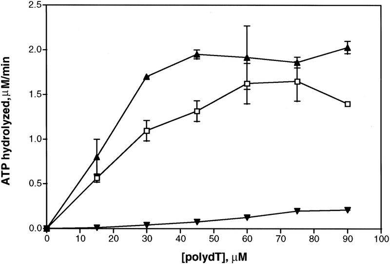
RadA protein is a DNA-dependent ATPase. This activity saturates at an apparent stoichiometry of 1 RadA monomer per 3 nucleotides at 75°C. The error bars indicate the s.d. for three sets of experiments. (▴) 15 μm RadA at 75°C; (□) 15μm RadA at 65°C; (▾) 15 μm RadA at 37°C.
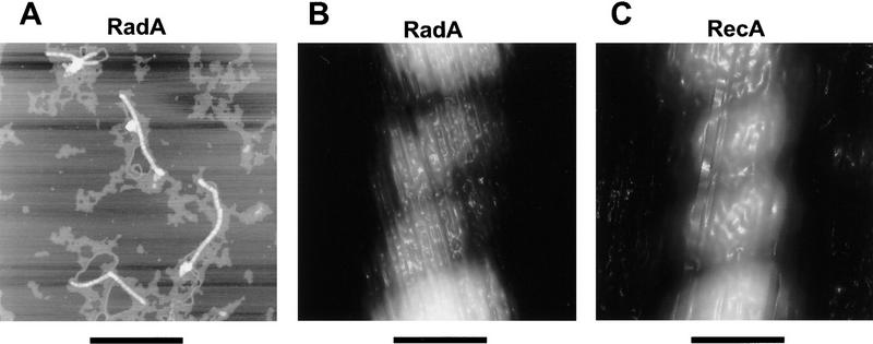
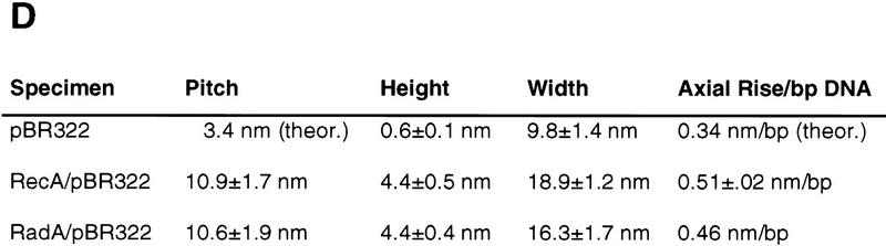
(A) Complexes of RadA protein and pBR322 plasmid DNA imaged by AFM. Bar, 500 nm. (B) The RadA protein-dsDNA filament is a right-handed helical structure that is similar to the RecA protein–dsDNA filament shown in C. Bars (B and C), 10 nm. (D) Physical dimensions of the RecA and RadA nucleoprotein filaments. The axial rise per bp for RadA/pBR322 is based on one measurement. The value reported for RecA/pBR322 is based on three measurements. Reported values for pitch, height, and width are each based on at least 10 measurements.
(A) Complexes of RadA protein and pBR322 plasmid DNA imaged by AFM. Bar, 500 nm. (B) The RadA protein-dsDNA filament is a right-handed helical structure that is similar to the RecA protein–dsDNA filament shown in C. Bars (B and C), 10 nm. (D) Physical dimensions of the RecA and RadA nucleoprotein filaments. The axial rise per bp for RadA/pBR322 is based on one measurement. The value reported for RecA/pBR322 is based on three measurements. Reported values for pitch, height, and width are each based on at least 10 measurements.

RadA protein can promote the formation of joint molecules. (A) DNA substrates and the expected product of the reaction. (B) An agarose gel displaying a time course for formation of joint molecules. The first six lanes show RadA protein-dependent D-loop formation with the 54-mer oligonucleotide SKBT17 at 65°C; the second six lanes show RadA protein-dependent D-loop formation with the 54-mer SKBT16 at 65°C; the third six lanes show the same reaction with SKBT16 in the absence of protein at 65°C; the last two lanes show RecA protein-dependent D-loop formation with SKBT16 at 37°C. (C) Joint molecule formation is dependent on RadA protein and DNA sequence homology. The error bars indicate the s.d. for three sets of experiments. (▪) SKBT16; (▵) SKBT17; (▾) PBR322 heterologous control; (⋄) no RadA.
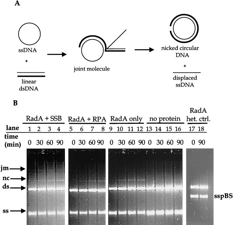
RadA protein promotes DNA pairing and strand exchange. (A) The DNA substrates as well as the expected intermediates and products of the DNA strand exchange reaction. (B) An agarose gel displaying a time course for formation of these products. Viral φX174 ssDNA (ss) and linear φX174 dsDNA (ds) were incubated with RadA protein, either with or without a ssDNA binding protein in lanes 1–12. Lanes 13–16 show the same reaction without RadA protein; lanes 17 and 18 show the same reaction with heterologous DNA. (jm) Joint molecules; (nc) nicked circular dsDNA; (sspBS) pBluescript ssDNA used as a heterologous control.
References
-
- Bult CJ, White O, Olsen GJ, Zhou L, Fleischmann RD, Sutton GG, Blake JA, Fitzgerald LM, Clayton RA, Gocayne JD. Complete genome sequence of the methanogenic archaeon, Methanococcus jannaschii. Science. 1996;273:1058–1073. - PubMed
-
- Bustamante C, Vesenka J, Tang CL, Rees W, Guthold M, Keller R. Circular DNA molecules imaged in air by scanning force microscopy. Biochemistry. 1992;31:22–26. - PubMed
-
- Chédin, F., E.M. Seitz, and S.C. Kowalczykowski. 1998. A highly conserved DNA-binding motif permits identification of a replication protein-A homologue in Archaea. Trends Biochem. Sci. (in press). - PubMed
-
- Cox MM. Binding of two DNA molecules at once: The recA protein. In: Revzin A, editor. The biology of nonspecific DNA-protein interactions. Boca Raton, FL: CRC Press; 1990. pp. 171–196.
Publication types
MeSH terms
Substances
Grants and funding
LinkOut - more resources
Full Text Sources
Other Literature Sources
Molecular Biology Databases
Research Materials