. 1998 May 1;12(9):1290-303.
doi: 10.1101/gad.12.9.1290.

Control of distal antennal identity and tarsal development in Drosophila by spineless-aristapedia, a homolog of the mammalian dioxin receptor

Affiliations

Control of distal antennal identity and tarsal development in Drosophila by spineless-aristapedia, a homolog of the mammalian dioxin receptor

D M Duncan et al. Genes Dev. .

Abstract

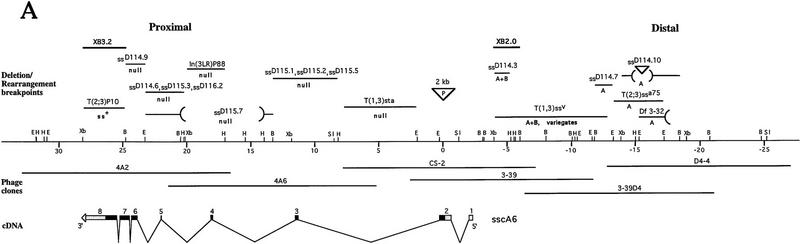
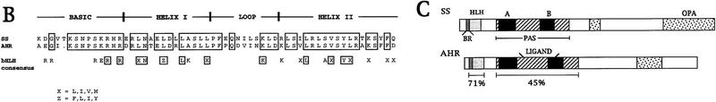
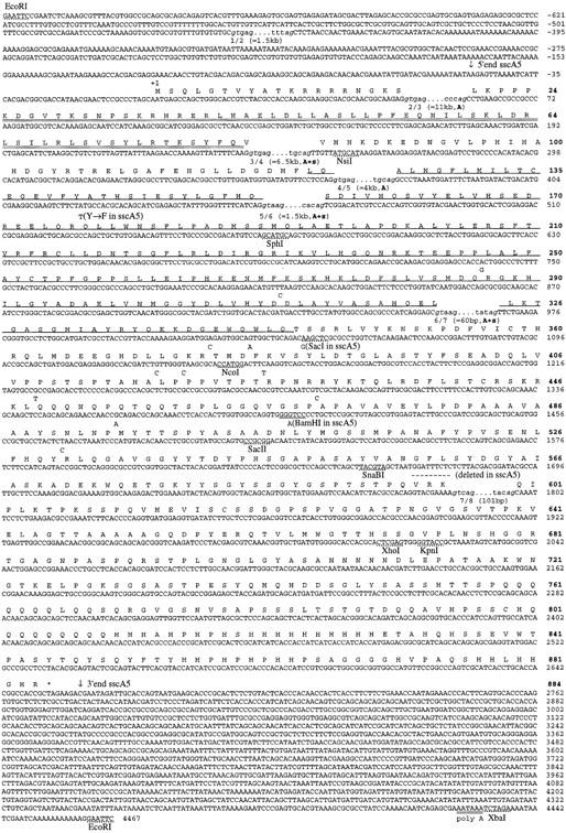
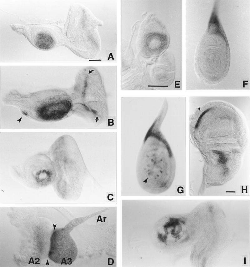
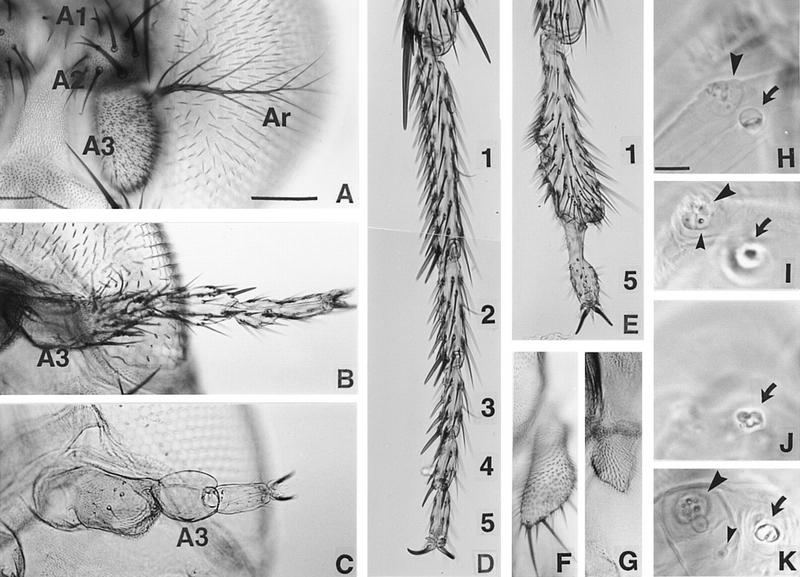
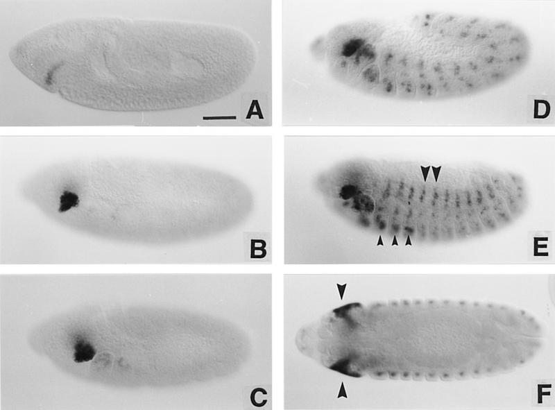
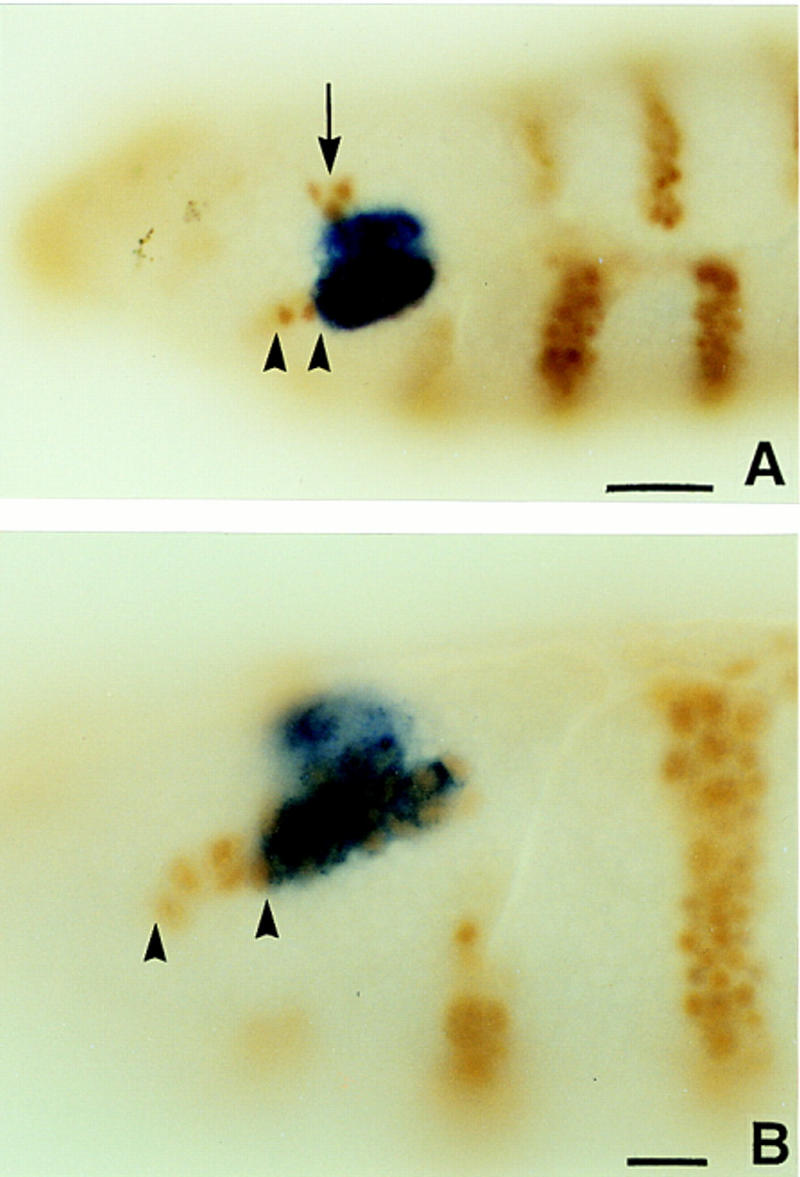
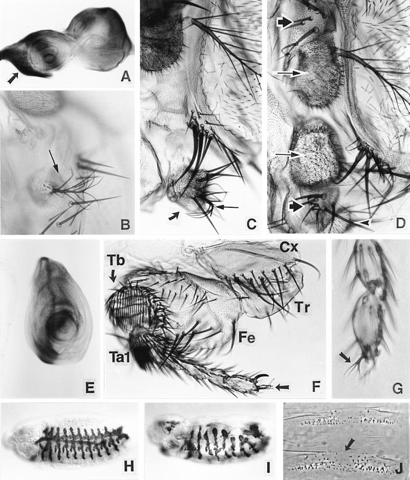
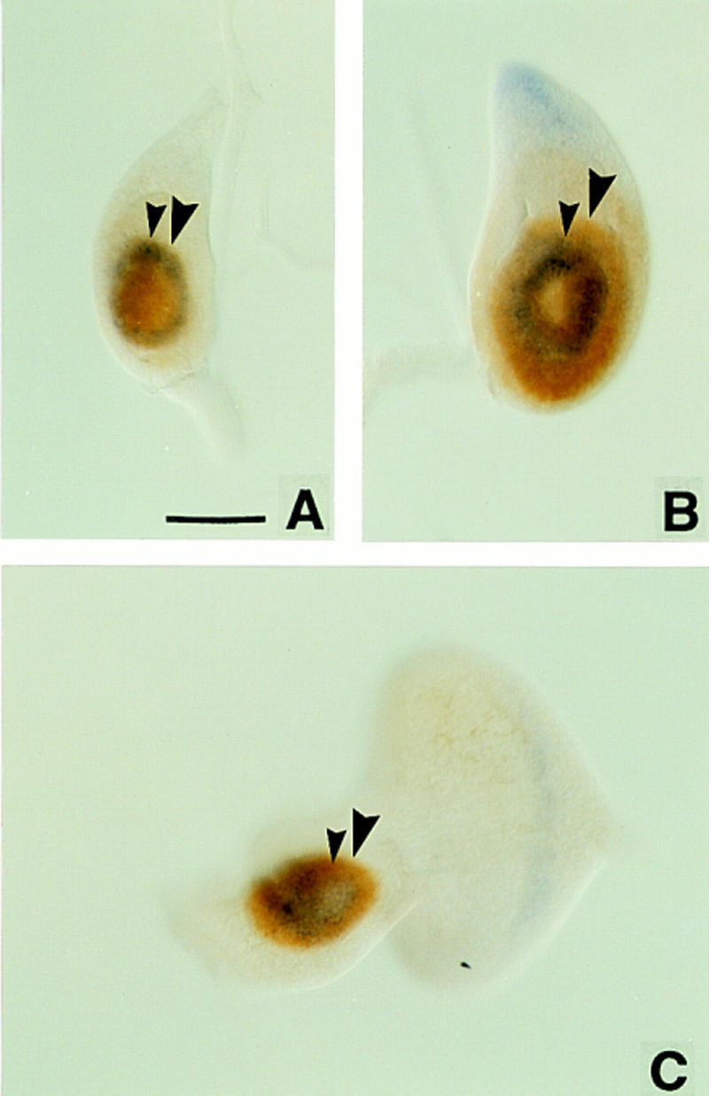
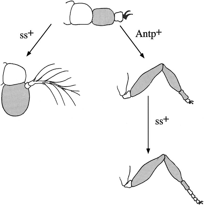
We report the molecular characterization of the spineless (ss) gene of Drosophila, and present evidence that it plays a central role in defining the distal regions of both the antenna and leg. ss encodes the closest known homolog of the mammalian dioxin receptor, a transcription factor of the bHLH-PAS family. Loss-of-function alleles of ss cause three major phenotypes: transformation of distal antenna to leg, deletion of distal leg (tarsal) structures, and reduction in size of most bristles. Consistent with these phenotypes, ss is expressed in the distal portion of the antennal imaginal disc, the tarsal region of each leg disc, and in bristle precursor cells. Ectopic expression of ss causes transformation of the maxillary palp and distal leg to distal antenna, and induces formation of an ectopic antenna in the rostral membrane. These effects indicate that ss plays a primary role in specifying distal antennal identity. In the tarsus, ss is expressed only early, and is required for later expression of the tarsal gene bric à brac (bab). Ectopic expression causes the deletion of medial leg structures, suggesting that ss plays an instructive role in the establishment of the tarsal primordium. In both the antenna and leg, ss expression is shown to depend on Distal-less (Dll), a master regulator of ventral appendage formation. The antennal transformation and tarsal deletions caused by ss loss-of-function mutations are probably atavistic, suggesting that ss played a central role in the evolution of distal structures in arthropod limbs.


Figures

Figure 1
Figure 1
Molecular analysis of the ss locus. (A) Map of the cloned DNA. Coordinates are in kb; position 0 is the insertion site of the P-element tag used to clone the locus. (B) BamHI; (E) EcoRI; (H) HindIII; (Sl) SalI; (Xb) XbaI. Overlapping phage clones are indicated below the restriction map; phage 3-39 is the P-element containing clone that initiated the walk. The positions of 17 breakpoint mutants are noted above the map; breakpoints and their uncertainties are indicated by lines, deletions by parentheses, and insertions by inverted triangles. The phenotypes of these mutants are noted as null, antennal transformations (A) or antennal transformations coupled with bristle defects (A+B). XB3.2 and XB2.0 indicate genomic fragments that detect transcripts when used to probe Northern blots. Coding sequences and untranslated regions within the exons of the sscA6 cDNA are indicated by black and shaded boxes, respectively. (B) Amino acid identity of ss to AHR in the bHLH domain. Boxed amino acids are identical between the two proteins, and identities of both ss and AHR to the bHLH consensus is noted below. (C) Structure of the ss and AHR proteins drawn to scale. Motifs are noted on the ss protein schematic. (BR) Basic region; (HLH) helix–loop–helix; (A, B, PAS) A and B repeats of the PAS domain; (OPA) region of opa or CAX repeats. The position of the ligand binding domain of AHR is also noted.
Figure 1
Figure 1
Molecular analysis of the ss locus. (A) Map of the cloned DNA. Coordinates are in kb; position 0 is the insertion site of the P-element tag used to clone the locus. (B) BamHI; (E) EcoRI; (H) HindIII; (Sl) SalI; (Xb) XbaI. Overlapping phage clones are indicated below the restriction map; phage 3-39 is the P-element containing clone that initiated the walk. The positions of 17 breakpoint mutants are noted above the map; breakpoints and their uncertainties are indicated by lines, deletions by parentheses, and insertions by inverted triangles. The phenotypes of these mutants are noted as null, antennal transformations (A) or antennal transformations coupled with bristle defects (A+B). XB3.2 and XB2.0 indicate genomic fragments that detect transcripts when used to probe Northern blots. Coding sequences and untranslated regions within the exons of the sscA6 cDNA are indicated by black and shaded boxes, respectively. (B) Amino acid identity of ss to AHR in the bHLH domain. Boxed amino acids are identical between the two proteins, and identities of both ss and AHR to the bHLH consensus is noted below. (C) Structure of the ss and AHR proteins drawn to scale. Motifs are noted on the ss protein schematic. (BR) Basic region; (HLH) helix–loop–helix; (A, B, PAS) A and B repeats of the PAS domain; (OPA) region of opa or CAX repeats. The position of the ligand binding domain of AHR is also noted.
Figure 2
Figure 2
Sequence of the ss cDNAs. The sequence and conceptual translation of the ss cDNA sscA6 is shown, along with the polymorphisms associated with the sscA5 cDNA. The nucleotide sequence is numbered by plain text at the end of each row; the corresponding amino acid sequence numbering is depicted in boldface type. Downward-pointing arrows indicate the ends of the sscA5 cDNA; broken lines above nucleotides +1789 through +1797 indicate the three codons absent in the sscA5 cDNA. The bHLH region is double underlined; the PAS domain is single underlined. The translation stop is noted by an asterisk. The positions of the seven exon splice junctions are in lowercase text; the donor and splice acceptors are italicized; the three flanking intronic nucleotides are in plain text. The approximate length of each intron is in parentheses, and those splice sites conserved in Ahr or sim are noted by a bold A and s, respectively.
Figure 3
Figure 3
ss expression in imaginal discs. Imaginal discs from third instar larvae and pupae were hybridized with RNA probes transcribed from the XB3.2 genomic clone or the sscA6 cDNA. (A,B) Eye–antennal discs from early and late wild-type third instar larvae. In B the arrowhead indicates the maxillary palp anlage and the arrows indicate the morphogenetic furrow in the eye disc. (C) ss transcript accumulation in ssa mutant (ssD114.3/ssD114.4) third instar eye–antennal discs. The pattern resembles that in the wild-type early third instar leg (cf. E). (D) ss expression in an everted antennal disc. Arrowheads indicate the boundary between the second (A2) and third (A3) segments. (Ar) The arista. (E– G) ss expression pattern in early E and late F third instar leg discs, and in a leg disc just prior to eversion (G). The arrowhead in G indicates bristle precursor cell labeling. (H) ss expression in a mature third instar wing disc. Expression is seen in an anteroventral stripe (arrowhead) and a patch in the presumptive notal region. (I) ss antennal expression in a heterozygote for the Antp gain-of-function allele Antp73b. Variegated reduction in ss expression is seen. Scale bars, 50 μm; the bar in A refers to A– D and I; the bar in E refers to E, F, and G.
Figure 4
Figure 4
Cuticular phenotypes of ss mutants. The antennal and leg phenotypes of ss mutants have been described by others [see Lindsley and Zimm (1992) and references therein]. (A,B) Antennae from wild-type (A) and ssa (ssD114.10/ssD114.4) (B) adults. The first (A1), second (A2), and third (A3) antennal segments and the arista (Ar) are indicated. The antennal tarsus in ssa mutants is judged to have T2 identity by the following criteria: Paired rows of stout bracted macrochaetae present ventrally (Hollingsworth 1964) are like those on the second leg; the most distal bristle pair in each segment is larger than more proximal pairs; on average, there are three pairs of bristles in the fifth tarsal segment (Lawrence et al. 1979); and there are no posterior transverse bristle rows in the second tarsal segment. (C) Antenna from a ss null mutant (ssD115.7). A3 has no bristles or trichomes, and most of the distal tarsal region is absent. (D–E) Distal second leg of wild-type and a ss null mutant. Tarsal segments 2–4 and part of segment 1 are deleted in E. (F,G) Wild-type and ss null mutant maxillary palps. The mutant palps are truncated. (H– K) Wild-type (H,I) and ss null mutant (J,K) first instar antennal and maxillary sense organs (AnSO and MxSO). The AnSO is indicated with a large arrow, the MxSO with a large arrowhead, and the dorsomedial papilla (DMP) with a small arrowhead. In the mutant the AnSO is cauliflower-shaped and has no clear stalk, and migration of the DMP is impaired. Scale bars, A–G (shown in A), 100 μm; H–K (shown in H), 10 μm.
Figure 5
Figure 5
ss transcript distribution in embryos. (A– E) The pattern of ss transcript distribution beginning at stage 8 and continuing through late embryogenesis. Small arrowheads in E indicate the leg anlage; large arrowheads indicate expression in the peripheral nervous system. In F, an optical section midway through a germ-band retracted embryo shows the extent of expression in the invaginating eye–antennal discs (arrowheads). Scale bar, 50 μm.
Figure 6
Figure 6
ss expression in the antennal segment of the embryo. (A,B) Wild-type embryos double labeled for ss transcript (blue) and en protein (brown). Note that the limits of ss expression extend from just ventral to the en head spot (arrow) through the antennal en stripe (arrowheads). The most ventral cells of the latter do not express ss. Scale bar in A, 20 μm; in B, 10 μm.
Figure 7
Figure 7
Effects of ss ectopic expression. (A) Pattern of ectopic expression driven by ptc–GAL4 in the eye–antennal disc (β-galactosidase staining). The arrow indicates a zone of high-level ectopic expression that includes the primordia of the palp and rostral membrane. (B–D) The effects of ectopic expression of ss driven by ptc–GAL4 in the head. A partial ectopic antenna (arrow) that has developed in an out pocketing of the rostral membrane is shown in B, and partial transformation of the palp (thick arrow) to arista (thin arrow) and AIII is shown in C. In D, an almost complete ectopic antenna is present in the rostral membrane region. Note that AIII (long arrows) and AII (short arrows) in the normal and ectopic antennae are arranged mirror symmetrically. The palp (arrowhead) is still present. The arista of the ectopic antenna is out of the plane of focus. (E– G) The effects of ectopic ss in the leg. The pattern of ectopic expression driven in the leg disc by ptc–GAL4 is shown in E. Deletion of most of the femur (Fe) and tibia (Tb) is shown in F. The coxa (Cx), trochanter (Tr), and most of the tarsus (Ta) are unaffected. Aristae are present at the distal tips of the legs shown in F and G (see arrows). (H–J) Effects of ectopic ss in embryos. H shows a normal embryo stained for the trh enhancer trap 1-eve-1 (Wilk et al. 1996). Ectopic ss driven by the 69B GAL4 driver causes severe abnormalities in this pattern (I). Ectopic ss also causes the deletion of midline portions of the denticle belts (arrow in J), similar to the effects of sim mutants.
Figure 8
Figure 8
Wild-type imaginal discs double-labeled for ss transcript (blue) and Dll protein (brown). Small arrowheads indicate the proximal extent of ss expression; large arrowheads indicate the proximal extent of Dll expression. In early third instar leg discs (A), the proximal extents of ss and Dll expression coincide; later discs show Dll expression more proximal than ss (B). In antennal discs, Dll expression is always more proximal than ss (C). Scale bar, 50 μm.
Figure 9
Figure 9
The ss antennal appendage is close to an epigenetic ground state. The antennal appendage in ss animals is indicated schematically at the top. The third segment of this appendage and its derivatives in the antenna and leg are shaded to indicate homology. As indicated, ss+ functions in the antenna to specialize the third and fourth segments of the ground state appendage to produce AIII and arista. In T2, Antp+ specifies the identities of the proximal two segments of the ground state appendage as coxa and trochanter, and directs the third segment to expand and subsegment to produce the femur, tibia, and first tarsal segment. These functions of Antp are consistent with the phenotype of Antp clones in the leg (Struhl 1981, 1982b; Abbott and Kaufman 1986), and with the effects of ectopic expression of Antp in the antenna (Postlethwait and Schneiderman 1971). In the distal leg, ss+ directs the expansion and segmentation of the tarsus. The diagram predicts that the Antp ss second leg will look like the ss antennal appendage. Although this has not yet been tested, the predicted phenotype is consistent with the additive effects of ss and Antp single mutants. The proximal two segments of the indicated ground state appendage are normal antennal segments; presumably the identities of these are specified by some other, currently unknown, homeotic gene or genes. Whether the evolutionary ground state of the limb is the same as the developmental ground state is not clear. However, it seems reasonable to suggest that it may be, and that an ancestral four-segmented appendage similar to the ss antenna became specialized in the head to produce the antenna, and in the thorax to produce legs.

References

    1. Abbot MK, Kaufman TC. The relationship between the functional complexity and the molecular organization of the Antennapedia locus of Drosophila melanogaster. Genetics. 1986;114:919–942. - PMC - PubMed
    1. Altschul SF, Gish W, Miller W, Myers EW, Lipman DJ. Basic local alignment search tool. J Mol Biol. 1990;215:403–410. - PubMed
    1. Baylies MK, Martinez Arias A, Bate M. wingless is required for the formation of a subset of muscle founder cells during Drosophila embryogenesis. Development. 1995;121:3829–3837. - PubMed
    1. Boudreaux HB. Arthropod Phylogeny, with special reference to insects. Malabar, FL: Robert E. Krieger Publishing; 1987.
    1. Brand A, Perrimon N. Targeted gene expression as a means of altering cell fates and generating dominant phenotypes. Development. 1993;118:401–415. - PubMed

MeSH terms

Associated data

LinkOut - more resources