. 1998 May 1;12(9):1327-37.
doi: 10.1101/gad.12.9.1327.

A ribosomal function is necessary for efficient splicing of the T4 phage thymidylate synthase intron in vivo

Affiliations

A ribosomal function is necessary for efficient splicing of the T4 phage thymidylate synthase intron in vivo

K Semrad et al. Genes Dev. .

Abstract

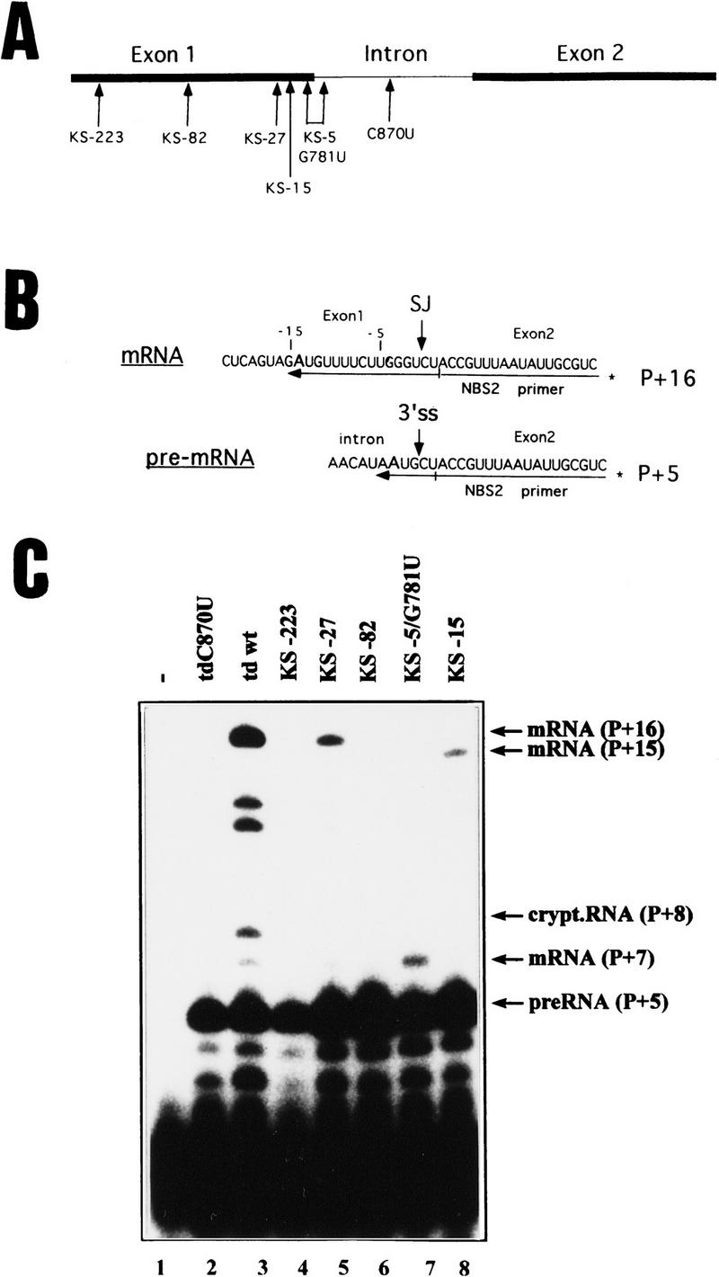
Splicing of the group I intron of the T4 thymidylate synthase (td) gene was uncoupled from translation by introducing stop codons in the upstream exon. This resulted in severe splicing deficiency in vivo. Overexpression of a UGA suppressor tRNA partially rescued splicing, suggesting that this in vitro self-splicing intron requires translation for splicing in vivo. Inhibition of translation by the antibiotics chloramphenicol and spectinomycin also resulted in splicing deficiency. Ribosomal protein S12, a protein with RNA chaperone activity, and CYT-18, a protein that stabilizes the three-dimensional structure of group I introns, efficiently rescued the stop codon mutants. We identified a region in the upstream exon that interferes with splicing. Point mutations in this region efficiently alleviate the effect of a nonsense codon. We infer from these results that the ribosome acts as an RNA chaperone to facilitate proper folding of the intron.


Figures

Figure 1
Figure 1
In vivo splicing of the stop codon mutants. (A) Position of the stop codons introduced into exon 1 of the T4 phage td gene and of the C870U intron mutant. Numbering of nucleotides indicates the distance to the 5′ splice site. For exact sequence of mutations see Table 1. (Solid bars) Exons; (thin lines) intron sequences. (B) Schematic representation of the primer–extension assay for splicing in E. coli. Extension of the 32P-labeled NBS2 primer, which hybridizes to exon 2 sequences, stops at the first A of the template in the presence of dATP, dCTP, dGTP and high concentrations of ddTTP. (P + x) Length of the extension product. pre-RNA is extended by 5 nucleotides (P + 5) and mRNA is extended by 16 nucleotides (P + 16). (SJ) Spliced junction; (3′ ss) 3′ splice site. (C) Primer-extension assay for splicing. The RNAs were isolated from cells containing no td gene (lane 1), the splicing-deficient mutant (td C870U, lane 2), the wild-type gene (lane 3), or the stop codon mutants as indicated (lanes 4–8). For the stop codon mutants KS-15 and KS-5/G781U the reverse transcripts of the mRNA differ in length because of the U to A mutation at position −15 or at position −5 and result in P + 15 and P + 7 extension products, respectively.
Figure 2
Figure 2
Stability of a nonsense codon containing mRNA. Comparison of the mRNA stability of the intronless mutants without and with a stop codon at position −82. The intronless constructs were grown to exponential phase and samples were taken before the addition of rifampicin (0′) and after its addition (1′ to 40′). mRNA amounts were assayed via primer-extension assay. (Lane 1) C870U mutant; (lane 2) wild type; (lanes 3–8) intronless td gene; (lanes 9–14) intronless td gene with stop codon at position −82.
Figure 3
Figure 3
Splicing of td mutants affecting the UAA stop codon at the 5′ site and the reading frame of exon 1. (A) The UAA stop codon, which immediately follows the 5′ splice site was mutated to UGA and UAG or readthrough (tdA771U). The amino acid sequence of the exon 1 product was modified via deletion of nucleotide −182 and insertion of a C at position −18, resulting in a product with 55 altered amino acids. (B) Splicing activity. The RNAs were isolated from E. coli cells containing no td gene (lane 1), the mutant tdC870U (lane 2), the wild type (lane 3) or the mutants described (lanes 5–9). The bands referring to mRNA, cryptic RNA and precursor are indicated by arrows. (C) P1 and cryptic P1. (P1) Sequences around the 5′ splice site (5′ ss) pair with intron sequences forming the P1 stem. Exon sequences are in uppercase, intron sequences in lowercase letters. The UAA stop codon following the 5′ ss is boxed. Positions altering this stop codon are indicated by arrows and the exchanged nucleotide is indicated. (Cryptic P1) An alternative folding of sequences surrounding the 5′ splice site is shown (Chandry and Belfort 1987). The original and the cryptic splice sites are indicated by arrows, as well as positions altered by mutations that stabilize the cryptic P1 folding. Negative numbers indicate distances to the 5′ splice site, positive numbers are intron positions.
Figure 3
Figure 3
Splicing of td mutants affecting the UAA stop codon at the 5′ site and the reading frame of exon 1. (A) The UAA stop codon, which immediately follows the 5′ splice site was mutated to UGA and UAG or readthrough (tdA771U). The amino acid sequence of the exon 1 product was modified via deletion of nucleotide −182 and insertion of a C at position −18, resulting in a product with 55 altered amino acids. (B) Splicing activity. The RNAs were isolated from E. coli cells containing no td gene (lane 1), the mutant tdC870U (lane 2), the wild type (lane 3) or the mutants described (lanes 5–9). The bands referring to mRNA, cryptic RNA and precursor are indicated by arrows. (C) P1 and cryptic P1. (P1) Sequences around the 5′ splice site (5′ ss) pair with intron sequences forming the P1 stem. Exon sequences are in uppercase, intron sequences in lowercase letters. The UAA stop codon following the 5′ ss is boxed. Positions altering this stop codon are indicated by arrows and the exchanged nucleotide is indicated. (Cryptic P1) An alternative folding of sequences surrounding the 5′ splice site is shown (Chandry and Belfort 1987). The original and the cryptic splice sites are indicated by arrows, as well as positions altered by mutations that stabilize the cryptic P1 folding. Negative numbers indicate distances to the 5′ splice site, positive numbers are intron positions.
Figure 4
Figure 4
Splicing inhibition by translation inhibitors. Splicing activity was tested in the absence and in the presence of increasing amounts of the antibiotics chloramphenicol and spectinomycin. (Lanes 1, 6) In vivo isolated wild-type RNA grown without antibiotic. (Lanes 2–5, 7–10) RNA isolated from the strains grown in the presence of increasing amounts (8, 40, 80 and 123 μm) of chloramphenicol and spectinomycin, respectively. Bands are as indicated in Fig. 1.
Figure 5
Figure 5
A UGA suppressor tRNA partially rescues splicing in a UGA mutant. Mutant KS-82 was cotransformed with a compatible plasmid encoding a UGA supressor tRNA. (A) In vivo RNA was isolated from the KS -82 mutant alone and from the strain cotransformed with the supressor tRNA on a plasmid, as described in the legend to Fig. 1. As controls the extension products from the wild type and the splicing-deficient mutant (td C870U) are shown. (B) The strains (as indicated) were plated on a thymine–deficient medium to test the suppressor activity and the effect of S12 overexpression on translation. Thy is thymine-deficient medium and Thy+ is a full medium.
Figure 6
Figure 6
Splicing rescue by ribosomal protein S12 and by CYT-18. The td wild-type and the stop codon mutants were cotransformed with a compatible plasmid encoding ribosomal protein S12 (A) and CYT 18 (B). In vivo splicing activity was tested via the primer-extension assay. The extension products are labeled as in Fig. 1.
Figure 6
Figure 6
Splicing rescue by ribosomal protein S12 and by CYT-18. The td wild-type and the stop codon mutants were cotransformed with a compatible plasmid encoding ribosomal protein S12 (A) and CYT 18 (B). In vivo splicing activity was tested via the primer-extension assay. The extension products are labeled as in Fig. 1.
Figure 7
Figure 7
Long-range interaction between exon 1 and the 3′ terminus of the intron. Potential base-pairing interaction between exon positions −81 to −73 and 3′-terminal sequences of the intron. Negative positions indicate distance to the 5′ splice site. Exon sequences are in uppercase, intron sequences in lowercase letters. (3′ ss) 3′ splice site. Mutations in mutant KS-82/1, which disrupt this pairing, are indicated. Underlined lowercase letters indicate intron positions involved in the three dimensional elements P9.0a and P9.0b (Jaeger et al. 1993; Michel et al. 1989).

Similar articles

Cited by

References

    1. Bacher G, Luetcke H, Jungnickel B, Rapoport TA, Dobberstein B. Regulation by the ribosome of the GTPase of the signal-recognition particle during protein targeting. Nature. 1996;381:248–251. - PubMed
    1. Belfort M, Moelleken A, Maley GF, Maley F. Purification and properties of T4 thymidylate synthase produced by the cloned gene in an amplification vector. J Biol Chem. 1983;258:2045–2051. - PubMed
    1. Belfort M, Chandry PS, Pedersen-Lane J. Genetic delineation of functional components of the group I intron in the phage T4 td gene. Cold Spring Harbor Symp Quant Biol. 1987;52:181–192. - PubMed
    1. Belfort M, Ehrenman K, Chandry PS. Genetic and molecular analysis of RNA splicing in Escherichia coli. Methods Enzymol. 1990;181:521–539. - PubMed
    1. Caprara MG, Lehnert V, Lambowitz AM, Westhof E. A tyrosyl-tRNA synthetase recognizes a conserved tRNA-like structural motif in the group I intron catalytic core. Cell. 1996a;87:1135–1145. - PubMed

Publication types

MeSH terms

LinkOut - more resources