. 1998 May 1;12(9):1338-47.
doi: 10.1101/gad.12.9.1338.

The ClpXP and ClpAP proteases degrade proteins with carboxy-terminal peptide tails added by the SsrA-tagging system

Affiliations

The ClpXP and ClpAP proteases degrade proteins with carboxy-terminal peptide tails added by the SsrA-tagging system

S Gottesman et al. Genes Dev. .

Abstract

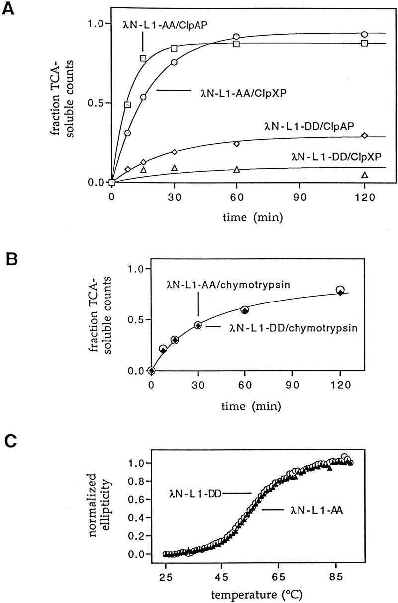
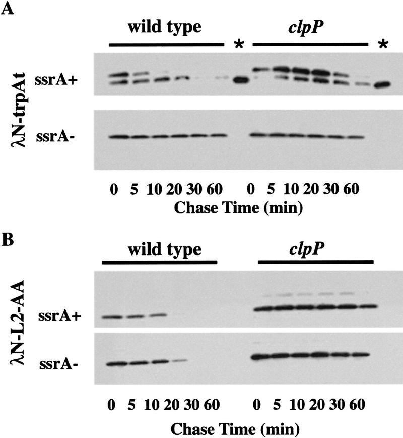
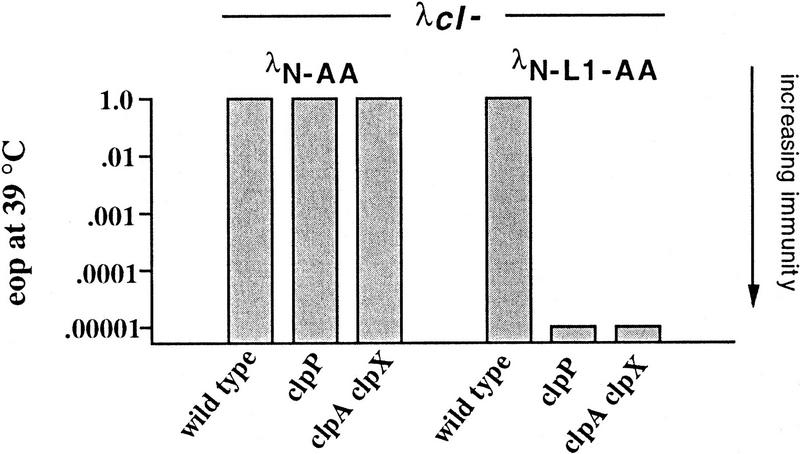
Interruption of translation in Escherichia coli can lead to the addition of an 11-residue carboxy-terminal peptide tail to the nascent chain. This modification is mediated by SsrA RNA (also called 10Sa RNA and tmRNA) and marks the tagged polypeptide for proteolysis. Degradation in vivo of lambda repressor amino-terminal domain variants bearing this carboxy-terminal SsrA peptide tag is shown here to depend on the cytoplasmic proteases ClpXP and ClpAP. Degradation in vitro of SsrA-tagged substrates was reproduced with purified components and required a substrate with a wild-type SsrA tail, the presence of both ClpP and either ClpA or ClpX, and ATP. Clp-dependent proteolysis accounts for most degradation of SsrA-tagged amino-domain substrates at 32 degrees C, but additional proteases contribute to the degradation of some of these SsrA-tagged substrates at 39 degrees C. The existence of multiple cytoplasmic proteases that function in SsrA quality-control surveillance suggests that the SsrA tag is designed to serve as a relatively promiscuous signal for proteolysis. Having diverse degradation systems able to recognize this tag may increase degradation capacity, permit degradation of a wide variety of different tagged proteins, or allow SsrA-tagged proteins to be degraded under different growth conditions.


Figures

Figure 1
Figure 1
Proteins used for degradation studies of SsrA-tagged proteins. Cartoon representation of the structures of hybrid proteins. The names of these proteins and of the plasmids that encode them are indicated to the left. [λ(1-93)] The 93-residue sequence of the amino-terminal, operator-binding domain of the λ cI repressor. (M2 Flag) The epitope sequence DYKDDDDK. (6His) The sequence HHHHHH. (SsrA tag AA) The sequence AANDENYALAA; (SsrA tag DD) The sequence AANDENYALDD. (trpAt) The AARLMSG sequence encoded by the stem–loop region of the trpAt transcriptional terminator. Note that because of heterogeneity in the position of SsrA tagging (Keiler et al. 1996), the λN–trpAt protein may consist of several species, some of which differ by 1 or 2 amino acids from the otherwise identical sequence of λN-L2-AA.
Figure 2
Figure 2
λN-AA and λN-L1-AA mediated immunity in clp mutants. Isogenic strains expressing λN-AA or λN-L1-AA were grown in tryptone broth with ampicillin and plated on LB–Amp plates in top agar. Ten-microliter serial dilutions of λcI or λc17 phage were spotted, plates were incubated overnight at 32°C, and an approximate eop was calculated relative to plating on the wild-type strain SG22163 transformed with pBR322. E. coli strains: wild-type (SG22163); clpP (SG22174); clpA (SG22176); clpX SG22177); and clpAclpX (SG22178). In control experiments, the heteroimmune phage λimm21cI, whose operators are not repressed by amino-terminal domain variants of λ repressor, plated with an eop of 1 on all strains containing λN-AA or λN-L1-AA and on wild-type strains expressing λN-DD or λN-L1-DD. λcI and λc17 plated with an eop of 10−4 or less on wild-type strains expressing λN-DD or λN-L1-DD.
Figure 3
Figure 3
Clp-dependent turnover in vivo of λN-AA and λN-L1-AA. (A) Western blot analysis of λN-AA in wild-type (SG22163) or clpP mutant (SG22175) cells after spectinomycin blocking of protein synthesis. Cells were grown to mid-log phase at 32°C and induced with IPTG for 30 min before addition of spectinomycin. Samples were removed at the times shown into cold TCA, precipitated, and resuspended for electrophoresis in 15% SDS–polyacrylamide gels. Gels were blotted and probed with anti-λ-repressor antibody. (B) Turnover of λN-L1-AA in wild-type and clp mutants. Experimental conditions were the same as in A except 10%–20% tricine SDS–polyacrylamide gels were used, and Western blots were probed with the M2 anti-FLAG antibody and quantified by the Eagle Eye II gel imaging system. Curves and strains are marked as for Fig. 2.
Figure 3
Figure 3
Clp-dependent turnover in vivo of λN-AA and λN-L1-AA. (A) Western blot analysis of λN-AA in wild-type (SG22163) or clpP mutant (SG22175) cells after spectinomycin blocking of protein synthesis. Cells were grown to mid-log phase at 32°C and induced with IPTG for 30 min before addition of spectinomycin. Samples were removed at the times shown into cold TCA, precipitated, and resuspended for electrophoresis in 15% SDS–polyacrylamide gels. Gels were blotted and probed with anti-λ-repressor antibody. (B) Turnover of λN-L1-AA in wild-type and clp mutants. Experimental conditions were the same as in A except 10%–20% tricine SDS–polyacrylamide gels were used, and Western blots were probed with the M2 anti-FLAG antibody and quantified by the Eagle Eye II gel imaging system. Curves and strains are marked as for Fig. 2.
Figure 4
Figure 4
Degradation in vitro of SsrA-tagged substrates by ClpAP and ClpXP. (A) Purified 35S-labeled λN-L1-AA or λN-L1-DD proteins were incubated with purified ClpAP or ClpXP in the presence of ATP at 30°C as described in Materials and Methods. Time-dependent release of TCA soluble counts is shown. (B) 35S-Labeled λN-L1-AA or λN-L1-DD were incubated with chymotrypsin using the same experimental conditions as in A. (C) Denaturation of the λN-L1-AA and λN-L1-DD proteins monitored by changes in circular dichroism shows no difference in thermal stability. Normalized CD ellipticity was calculated as (ε25−ε)/(ε25−ε93) where e is the ellipticity at 230 nm at each temperature and ε25 and ε93 are the ellipticities at 230 nm at 25°C and 93°C, respectively.
Figure 5
Figure 5
Degradation of λN-trpAt and λN-L2-AA in vivo. (A) Western blots of λN–trpAt turnover following spectinomycin treatment in ssrA+ clp+ (SG22163), ssrA+ clpP::kan (SG22175), ssrA::cat clp+ (SG22183), and ssrA::cat clpP::kan (SG22184) strains. Note the presence of tagged and untagged variants of the λN-trpAt protein in ssrA+ strains. Lanes marked with an asterisk are untagged samples from the ssrA host run on the same gel as ssrA+ samples to allow comparison of electrophoretic mobilities. Experimental procedures were the same as described in Fig. 3; 10%–20% gradient tricine gels (Novex) were used for electrophoresis. (B) Western blots of λN-L2-AA turnover. Strains and conditions were identical to those shown in A. In control experiments, λN-L2-DD expressed in the wild-type strain (SG22163) showed no significant turnover over 1 hr (data not shown). (C) Time courses of degradation of SsrA-tagged λN–trpAt protein (upper band in A) and λN-L2-AA in ssrA+ clp+ (SG22163) and ssrA+ clpP::kan (SG22175) strains. These data were obtained by scanning of the gels shown in A and B.
Figure 5
Figure 5
Degradation of λN-trpAt and λN-L2-AA in vivo. (A) Western blots of λN–trpAt turnover following spectinomycin treatment in ssrA+ clp+ (SG22163), ssrA+ clpP::kan (SG22175), ssrA::cat clp+ (SG22183), and ssrA::cat clpP::kan (SG22184) strains. Note the presence of tagged and untagged variants of the λN-trpAt protein in ssrA+ strains. Lanes marked with an asterisk are untagged samples from the ssrA host run on the same gel as ssrA+ samples to allow comparison of electrophoretic mobilities. Experimental procedures were the same as described in Fig. 3; 10%–20% gradient tricine gels (Novex) were used for electrophoresis. (B) Western blots of λN-L2-AA turnover. Strains and conditions were identical to those shown in A. In control experiments, λN-L2-DD expressed in the wild-type strain (SG22163) showed no significant turnover over 1 hr (data not shown). (C) Time courses of degradation of SsrA-tagged λN–trpAt protein (upper band in A) and λN-L2-AA in ssrA+ clp+ (SG22163) and ssrA+ clpP::kan (SG22175) strains. These data were obtained by scanning of the gels shown in A and B.
Figure 6
Figure 6
Phage immunity at 39°C. Immunity to λcI of wild-type (SG22163), clpP (SG22174), and clpA clpX (SG22178) strains expressing λN-AA and λN-AA at 39°C. Except for temperature, the plating conditions were identical to those described in Fig. 2.

References

    1. Benyon RJ, Bond JS. Proteolytic enzymes: A practical approach. Oxford, UK: IRL Press; 1989.
    1. Bowie JU, Sauer RT. Identification of C-terminal extensions that protect proteins from intracellular proteolysis. J Biol Chem. 1989;264:7596–7602. - PubMed
    1. Clarke ND, Beamer LJ, Goldberg HR, Berkower C, Pabo CO. The DNA binding arm of λ repressor: Critical contacts from a flexible region. Science. 1991;254:267–270. - PubMed
    1. Davidson AR, Sauer RT. Folded proteins occur frequently in libraries of random amino acid sequences. Proc Natl Acad Sci. 1994;91:2146–2150. - PMC - PubMed
    1. Felden B, Himeno H, Muto A, McCutcheon JP, Atkins JF, Gesteland RF. Probing the structure of the Escherichia coli 10Sa RNA (tmRNA) RNA. 1997;3:89–103. - PMC - PubMed

Publication types

MeSH terms

LinkOut - more resources