. 1998 May 1;12(9):1371-80.
doi: 10.1101/gad.12.9.1371.

Prespore-specific gene expression in Bacillus subtilis is driven by sequestration of SpoIIE phosphatase to the prespore side of the asymmetric septum

Affiliations

Prespore-specific gene expression in Bacillus subtilis is driven by sequestration of SpoIIE phosphatase to the prespore side of the asymmetric septum

L J Wu et al. Genes Dev. .

Abstract

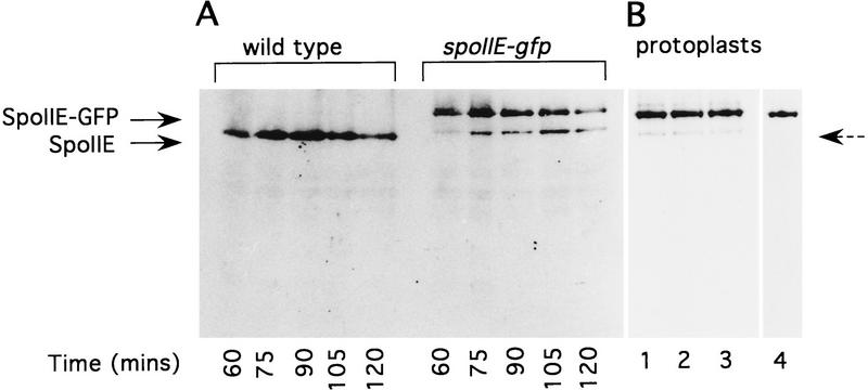
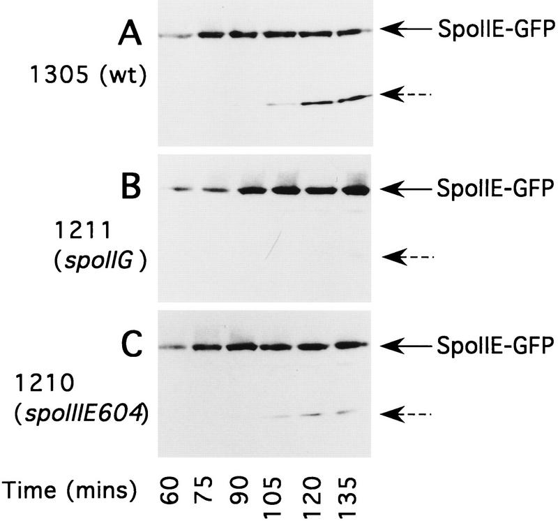
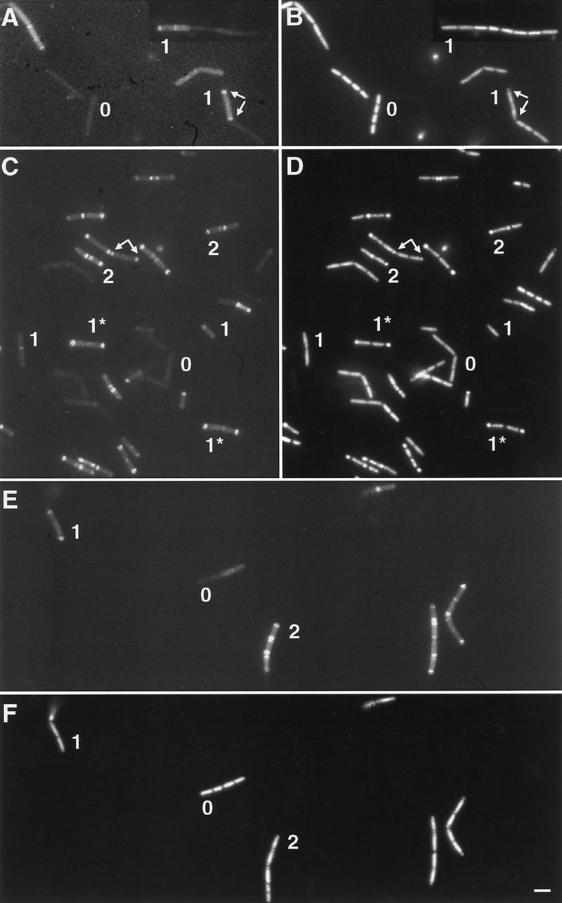
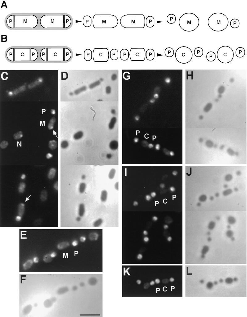
The spoIIE gene is essential for the compartment-specific activation of transcription factor sigmaF during sporulation in Bacillus subtilis. SpoIIE is a membrane protein that is targeted to the potential sites of asymmetric septation near each pole of the sporulating cell. The cytoplasmic carboxy-terminal domain of SpoIIE contains a serine phosphatase that triggers the release of sigmaF in the prespore compartment after septation. To understand how septum-located SpoIIE is activated selectively in the prespore, we examined the distribution of a SpoIIE-GFP fusion protein. We show that the polar bands of SpoIIE protein actually form sequentially and that the most prominent band develops at the pole where the prespore forms. We also show that the protein is sequestered to the prespore side of the asymmetric septum. Sequestration of SpoIIE into the prespore compartment provides a mechanism that could explain the cell specificity of sigmaF activation.


Figures

Figure 1
Figure 1
Accumulation and stability of SpoIIE and SpoIIE–GFP in sporulating cells and protoplasts. (A) Whole cell extracts of sporulating wild-type strain SG38 and the spoIIE–gfp-carrying strain 1305, taken at the times indicated, were analyzed by Western blot using anti-SpoIIE antibodies. (B) To demonstrate that protoplasting does not change the degradation pattern of SpoIIE–GFP, cells of strain 1305 were collected and protoplasted at t90, then treated in various ways before being subjected to Western blot analysis using anti-SpoIIE antibodies (lanes 1–3) or monoclonal anti-GFP antibody (lane 4). The protoplasts were treated as follows: (lane 1) pelleted and frozen immediately; left for 30 min (lanes 2,4) at room temperature or (lane 3) on ice, then pelleted and frozen. The positions of SpoIIE and SpoIIE–GFP are indicated by solid arrows. The broken arrow indicates the position of a faster migrating band that cross-reacted with anti-SpoIIE, possibly representing near full-length SpoIIE without the GFP tag.
Figure 2
Figure 2
Time course of SpoIIE/SpoIIE–GFP degradation during sporulation. Whole cell extracts from sporulating cells were loaded on a 10% SDS-polyacrylamide gel and analyzed by Western blot using monoclonal anti-GFP antibody. Samples were prepared from the following strains: (A) 1305 (spoIIE–gfp); (B) 1211(spoIIE–gfp spoIIGA::aph-A3); (C) 1210 (spoIIE–gfp spoIIIE604). Samples were taken and prepared as described in the legend to Fig. 1. The positions of full-length SpoIIE–GFP and the major SpoIIE–GFP degradation product are indicated by solid and broken arrows, respectively.
Figure 3
Figure 3
SpoIIE band formation and development. Strains carrying the spoIIE–gfp fusion were induced to sporulate, and live DAPI-stained cells were examined by fluorescence microscopy. (A,B) Wild-type (1305) cells at t70; (C,D) wild-type cells at t105; (E,F) spoIIG spoIIIE double mutant (strain 1213) at t90. (A,C,E) Localization of SpoIIE–GFP; (B,D,F) DAPI images showing the nucleoids of cells in the same fields as in A, C, and E, respectively. (0) Examples of cells without SpoIIE bands; (1) cells with one relatively weak and straight band, corresponding to cells that have not started to engulf; (1*) cells with one strong and curving fluorescent band, corresponding to cells undergoing or having completed engulfment; (2) cells with two SpoIIE bands. Arrows point to the asymmetric septa in cells that have not yet completed transfer of the prespore chromosome. Scale bar, 2.5 μm.
Figure 4
Figure 4
Localization of SpoIIE–GFP in protoplasted cells from sporulating cultures just before and after σF becomes active. (A,B) Schematic illustration of the consequences of treatment of (A) wild-type and (B) spoIIG mutant cells with lysozyme to make protoplasts. Removal of the cell wall material (shaded) allows the prespore (P), mother cell (M), or central (C) compartments to enlarge and round off and separate, allowing the fate of GFP initially located at the site of the thin division septum to be resolved. The remaining panels show images of typical cells. (C–F) Wild type (strain 1305); (G–J) spoIIG spoIIIE double mutant (strain 1213); (K,L) spoIIG mutant (strain 1211). (C,D,G,H) Cells at an early stage of sporulation (t85), just before activation of σF. (E,F,I–L) Cells at slightly later stages of sporulation (t100), just after σF becomes active. (C,E,G,I,K) Fluorescence images showing the distribution of SpoIIE–GFP; (D,F,H,J,L) phase-contrast images of same fields as C, E, G, I, and K, respectively. (N) Nonseptate cell. Arrows point to some of the SpoIIE spots in the mother cell protoplasts. Scale bar, 2.5 μm.
Figure 5
Figure 5
Absence of σF activity from the central compartment of a spoIIG spoIIIE double mutant. Various strains carrying a σF-dependent gpr–lacZ fusion located at the gpr locus (::pPS1395) or at amyE (::pPS1326) were induced to sporulate and β-galactosidase was assayed. (•) Wild-type strain SG38::pPS1395; (▴) spoIIG spoIIIE double mutant with the fusion located at the gpr locus (919::pPS1395); (▵) spoIIG spoIIIE double mutant with the fusion located at the amyE locus (919::pPS1326); (○) spoIIG single mutant 901::pPS1395; (□) spoIIIE single mutant 604.6::pSG1395.
Figure 6
Figure 6
Schematic summary of the localization of SpoIIE protein in wild-type and mutant cells (A) and possible models for sequestration of SpoIIE to the prespore compartment (B,C). (A) 1 represents a preseptational cell soon after the initiation of sporulation; open arrowheads indicate the potential sites for asymmetric septation close to each pole. σF is present throughout the cytoplasm but in an inactive state (indicated by outline text). SpoIIE protein (○) then begins to be made (2) and is localized initially in a band at the polar septation site where the asymmetric septum will be formed, presumably by association with a component of the division apparatus. When the septum has formed (3) the SpoIIE protein is sequestered specifically into the membrane on the prespore side, and a second band of SpoIIE starts to appear at the prespore-distal potential division site. Sequestration of most of the cellular SpoIIE into the small prespore compartment results in activation of σF (boldface type). In wild-type cells SpoIIE in the mother cell is then degraded (4), probably as a result of activation of σE and synthesis of one or more proteases encoded by genes controlled by the new form of RNA polymerase. In spoIIIE mutants (5), the second SpoIIE ring persists in the mother cell, leading to aberrant activation of σF in that compartment. In spoIIG single or spoIIG spoIIIE double mutant (6), the absence of σE allows the second polar septum to form. The second SpoIIE ring is thus sequestered into its prespore, leaving the central compartment devoid of SpoIIE, and hence preventing σF from being activated in this compartment. (B) SpoIIE protein localizes initially to the site of incipient septation. As the septal annulus closes, flux of membrane lipid from the large compartment (where the bulk of new lipid synthesis could conceivably take place) drives the membrane-bound SpoIIE protein into the prespore compartment. (C) The SpoIIE band forms just to the polar side of the division site, so that as the septum forms the protein is captured by the prespore.

Similar articles

Cited by

References

    1. Alper S, Duncan L, Losick R. An adenosine nucleotide switch controlling the activity of a cell type-specific transcription factor in B. subtilis. Cell. 1994;77:195–205. - PubMed
    1. Anagnostopoulos C, Spizizen J. Requirements for transformation in Bacillus subtilis. J Bacteriol. 1961;81:741–746. - PMC - PubMed
    1. Arigoni F, Pogliano K, Webb CD, Stragier P, Losick R. Localization of protein implicated in establishment of cell type to sites of asymmetric division. Science. 1995;270:637–640. - PubMed
    1. Arigoni F, Duncan L, Alper S, Losick R, Stragier P. SpoIIE governs the phosphorylation state of a protein regulating transcription factor σF during sporulation in Bacillus subtilis. Proc Natl Acad Sci. 1996;93:3238–3242. - PMC - PubMed
    1. Barak I, Youngman P. SpoIIE mutants of Bacillus subtilis comprise two distinct phenotypic classes consistent with a dual functional role for the SpoIIE protein. J Bacteriol. 1996;178:4984–4989. - PMC - PubMed

Publication types

MeSH terms

LinkOut - more resources