Immunization of cattle by infection with Cowdria ruminantium elicits T lymphocytes that recognize autologous, infected endothelial cells and monocytes
- PMID: 9573061
- PMCID: PMC108135
- DOI: 10.1128/IAI.66.5.1855-1860.1998
Immunization of cattle by infection with Cowdria ruminantium elicits T lymphocytes that recognize autologous, infected endothelial cells and monocytes
Abstract
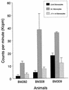
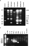
Peripheral blood mononuclear cells (PBMC) from immune cattle proliferate in the presence of autologous Cowdria ruminantium-infected endothelial cells and monocytes. Endothelial cells required treatment with T-cell growth factors to induce class II major histocompatibility complex expression prior to infection and use as stimulators. Proliferative responses to both infected autologous endothelial cells and monocytes were characterized by expansion of a mixture of CD4+, CD8+, and gammadelta T cells. However, gammadelta T cells dominated following several restimulations. Reverse transcription-PCR analysis of cytokine expression by C. ruminantium-specific T-cell lines and immune PBMC revealed weak interleukin-2 (IL-2), IL-4, and gamma interferon (IFN-gamma) transcripts at 3 to 24 h after stimulation. Strong expression of IFN-gamma, tumor necrosis factor alpha (TNF-alpha), TNF-beta, and IL-2 receptor alpha-chain mRNA was detected in T-cell lines 48 h after antigen stimulation. Supernatants from these T-cell cultures contained IFN-gamma protein. Our findings suggest that in immune cattle a C. ruminantium-specific T-cell response is induced and that infected endothelial cells and monocytes may present C. ruminantium antigens to specific T lymphocytes in vivo during infection and thereby play a role in induction of protective immune responses to the pathogen.
Figures


Similar articles
-
Identification of Cowdria ruminantium antigens that stimulate proliferation of lymphocytes from cattle immunized by infection and treatment or with inactivated organisms.Infect Immun. 2000 Feb;68(2):603-14. doi: 10.1128/IAI.68.2.603-614.2000. Infect Immun. 2000. PMID: 10639423 Free PMC article.
-
Immunisation of cattle against heartwater by infection with Cowdria ruminantium elicits T lymphocytes that recognise major antigenic proteins 1 and 2 of the agent.Vet Immunol Immunopathol. 2002 Feb;85(1-2):23-32. doi: 10.1016/s0165-2427(01)00421-4. Vet Immunol Immunopathol. 2002. PMID: 11867164
-
Cellular immune responses of cattle to Cowdria ruminantium.Dev Biol Stand. 1998;92:309-15. Dev Biol Stand. 1998. PMID: 9554286
-
Infection of bovine brain microvessel endothelial cells with Cowdria ruminantium elicits IL-1 beta, -6, and -8 mRNA production and expression of an unusual MHC class II DQ alpha transcript.J Immunol. 1995 Apr 15;154(8):4032-8. J Immunol. 1995. PMID: 7706742
-
Low molecular weight proteins of Cowdria ruminantium (Welgevonden isolate) induce bovine CD4+-enriched T-cells to proliferate and produce interferon-gamma.Vet Microbiol. 2002 Mar 22;85(3):259-73. doi: 10.1016/s0378-1135(01)00516-8. Vet Microbiol. 2002. PMID: 11852193
Cited by
-
Th1 and Th2 epitopes of Cowdria polymorphic gene 1 of Ehrlichia ruminantium.Onderstepoort J Vet Res. 2023 Mar 23;90(1):e1-e15. doi: 10.4102/ojvr.v90i1.2070. Onderstepoort J Vet Res. 2023. PMID: 37042556 Free PMC article.
-
Progress and obstacles in vaccine development for the ehrlichioses.Expert Rev Vaccines. 2010 Sep;9(9):1071-82. doi: 10.1586/erv.10.93. Expert Rev Vaccines. 2010. PMID: 20822349 Free PMC article. Review.
-
Kinetics of antibody response to Ehrlichia canis immunoreactive proteins.Infect Immun. 2003 May;71(5):2516-24. doi: 10.1128/IAI.71.5.2516-2524.2003. Infect Immun. 2003. PMID: 12704123 Free PMC article.
-
Identification of Cowdria ruminantium antigens that stimulate proliferation of lymphocytes from cattle immunized by infection and treatment or with inactivated organisms.Infect Immun. 2000 Feb;68(2):603-14. doi: 10.1128/IAI.68.2.603-614.2000. Infect Immun. 2000. PMID: 10639423 Free PMC article.
References
-
- Baldwin C L, Teale A J, Naessens J G, Goddeeris B M, MacHugh N D, Morrison W I. Characterization of a subset of bovine T lymphocytes that express BoT4 by monoclonal antibodies and function: similarity to lymphocytes defined by human T4 and murine L3T4. J Immunol. 1986;136:4385–4391. - PubMed
-
- Baldwin C L, Morrison W I, Naessens J. Differentiation antigens and functional characteristics of bovine leukocytes. In: Trnka Z, Miyasaka M, editors. Comparative aspects of differentiation antigens in lympho-haemopoietic tissues. New York, N.Y: Marcel Dekker, Inc.; 1988. pp. 455–498.
-
- Born W K, Harshan K, Modlin R L, O’Brien R. The role of γδ T lymphocytes in infection. Curr Opin Immunol. 1991;3:455–459. - PubMed
-
- Bourdoulous S, Bensaid A, Martinez D, Sheikboudou C, Trap I, Strosberg A, Couraud P. Infection of bovine brain microvessel endothelial cells with Cowdria ruminantium elicits IL-1β, -6, and -8 mRNA production and expression of unusual MHC class II DQα transcript. J Immunol. 1995;154:4032–4038. - PubMed
-
- Brown W C, Logan K S. Babesia bovis: bovine helper T cell lines reactive with soluble and membrane antigens of merozoites. Exp Parasitol. 1992;74:188–199. - PubMed
Publication types
MeSH terms
Substances
LinkOut - more resources
Full Text Sources
Other Literature Sources
Research Materials

