Comparative Study
. 1998 May;66(5):1918-27.
doi: 10.1128/IAI.66.5.1918-1927.1998.

Comparisons between colony phase variation of Neisseria gonorrhoeae FA1090 and pilus, pilin, and S-pilin expression

Affiliations
Comparative Study

Comparisons between colony phase variation of Neisseria gonorrhoeae FA1090 and pilus, pilin, and S-pilin expression

C D Long et al. Infect Immun. 1998 May.

Abstract

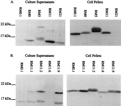
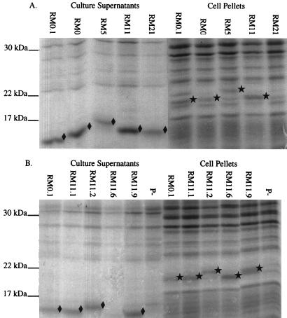
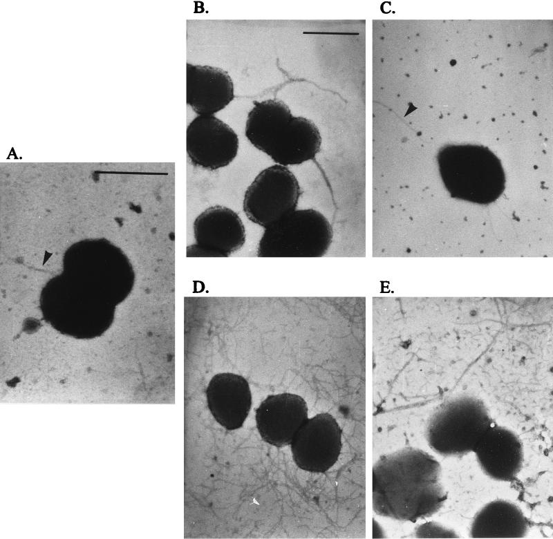
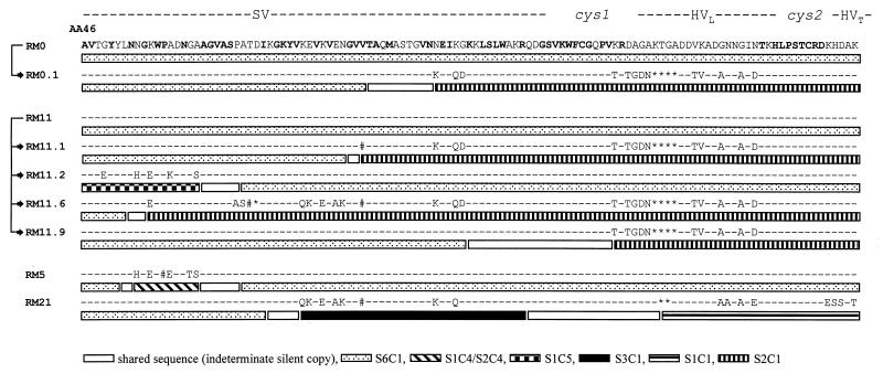
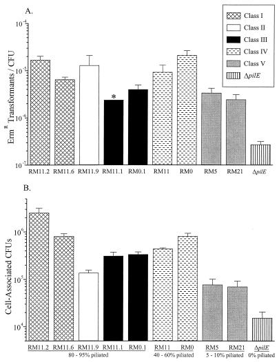
The gonococcal pilus is a primary virulence factor, providing the initial attachment of the bacterial cell to human mucosal tissues. Pilin, the major subunit of the pilus, can carry a wide spectrum of primary amino acid sequences which are generated by the action of a complex antigenic variation system. Changes in the pilin amino acid sequence can produce different pilus-dependent colony morphotypes, which have been previously shown to reflect phase variation of pili on the bacterial cell surface. In this study, we further examined the relationships between changes in pilus-dependent colony morphology, pilin sequence, pilus expression, and pilus function in Neisseria gonorrhoeae FA1090. A group of FA1090 colony variants expressed different pilin sequences and demonstrated different levels of pilin, S-pilin, and pilus expression. The analysis of these colony variants shows that they do not represent two distinct phases of pilus expression, but that changes in pilin protein sequence produce a spectrum of S-pilin production, pilus expression, and pilus aggregation levels. These different levels of pilus expression and aggregation influence not only colony morphology but also DNA transformation efficiency and epithelial cell adherence.


Figures

FIG. 1
FIG. 1
Pilin immunoblots of N. gonorrhoeae FA1090 colony variants. Soluble supernatants and cell pellets were separated on SDS-gradient polyacrylamide gels and probed with the broadly cross-reactive MAb 1E8/G8. (A) P+/− variants and P+ variant RM0.1 for comparison; (B) P+ variants. All variants contained the recA6 allele. RM0.1 S-pilin reacted poorly with MAb 1E8/G8 (A) but is detectable after a more extended development of the immunoblot (B).
FIG. 2
FIG. 2
Stained SDS-polyacrylamide gels of FA1090 colony variants. Soluble supernatants and cell pellets were separated on SDS-gradient polyacrylamide gels and stained with Coomassie brilliant blue. (A) P+/− variants and P+ variant RM0.1 for comparison; (B) P+ variants and a nonpiliated (ΔpilE) control (P). All P+ and P+/− variants contained the recA6 allele. S-pilin bands (⧫) and full-length pilin bands (★) as identified by parallel immunoblotting are marked.
FIG. 3
FIG. 3
Transmission electron micrographs of FA1090 colony variants. Representative examples of P+ cells are shown, but a combination of P+ and P cells was observed (Table 1). (A) Class V variant RM5 recA6; (B) class IV variant RM0 recA6; (C) class III variant RM11.1 recA6; (D) class I variant RM11.2 recA6; (E) class II variant RM11.9 recA6. Arrowheads in panels A and C mark pili. The bar in panel A represents 1 μm in panels A and C; the bar in panel B represents 1 μm in panels B, D, and E.
FIG. 4
FIG. 4
Predicted variable pilin amino acid sequences of FA1090 pilE variants. Variant names and lineages are shown on the left. DNA sequences were determined from PCR-amplified pilE DNA, but only the variable amino acid sequences are shown. The SV, cys1, HVL, cys2, and hypervariable tail (HVT) regions of pilE are indicated on the top. −, no changes relative to variant RM0; ∗, deletions relative to variant RM0; #, silent changes in DNA sequence not reflected in the amino acid sequence. Amino acid residues that have never varied in an expressed gene on any pilin variant from any reported N. gonorrhoeae strain are indicated in bold on the variant RM0 sequence. Patterned boxes represent nucleotide stretches of the pilE DNA sequences that can be mapped to a particular silent copy. The legend shows the silent locus and silent copy sources of each variable DNA sequence; for example, S1C1 indicates pilS1 copy 1 DNA sequences. Sequences at the junction of two different adjacent silent copy sequences that are identical in those silent copies are indicated as shared sequences; for example, RM0.1 has pilS6 copy 1-specific sequences and pilS2 copy 1-specific sequences, and the open box shows the sequence where these two silent copies are identical.
FIG. 5
FIG. 5
DNA transformation efficiency and epithelial cell adherence of FA1090 colony variants. All P+ and P+/− variants contained the recA6 allele. Values are mean ± standard error. Variants are grouped by piliation class as defined by TEM (Fig. 3). See also Table 1. (A) Average number of gonococci transformed to Ermr/total CFU. Three to nine experiments were performed per variant. ∗, the transformation frequency could not be accurately determined due to consistently low CFU, but is close to the value shown. (B) Average cell-associated N. gonorrhoeae CFU/well of subconfluent Chang conjunctival epithelial cell monolayers. Three to seven experiments were performed per variant.
FIG. 6
FIG. 6
Phenotypes of RM11.2 recA6 colony variants. To generate colony variants, RM11.2 recA6 was induced with 1 mM IPTG on plates for either 18 (A) or 24 (B) h. Colonies with a morphology less P+ than RM11.2 recA6 were categorized as either flat and without a distinct edge (P), or intermediate, lacking the dark ring found at the edge of most P+ colonies but more domed than P colonies (P+/−). The presence (pilE+), absence (ΔpilE), or larger size (L-pilin) of the pilE gene was determined by PCR analysis of colony lysates from 173 colony variants at 18 h and 210 colony variants at 24 h. The presence (pilin+) or absence (pilin) of pilin protein was determined by Western analysis of a subset of colonies from each time point (n = 32 at 18 h; n = 50 at 24 h). The percentage of colony variants representing true pilus phase variants is indicated by an arc (see Discussion).

Similar articles

Cited by

References

    1. Arminjon F, Cadoz M, Morse S A, Rock J P, Sarafian S K. Abstracts of the 87th Annual Meeting of the American Society for Microbiology 1987. Washington, D.C: American Society for Microbiology; 1987. Bactericidal and opsonic activities of sera from individuals immunized with a gonococcal protein I vaccine, abstr. E-92; p. 118.
    1. Barritt D S, Schwalbe R S, Klapper D G, Cannon J G. Antigenic and structural differences among six proteins II expressed by a single strain of Neisseria gonorrhoeae. Infect Immun. 1987;55:2026–2031. - PMC - PubMed
    1. Bergström S, Robbins K, Koomey J M, Swanson J. Piliation control mechanisms in Neisseria gonorrhoeae. Proc Natl Acad Sci USA. 1986;83:3890–3894. - PMC - PubMed
    1. Bessen D, Gotschlich E C. Interactions of gonococci with HeLa cells: attachment, detachment, replication, penetration, and the role of protein II. Infect Immun. 1986;54:154–160. - PMC - PubMed
    1. Bollag D M, Edelstein S J. Protein methods. New York, N.Y: John Wiley & Sons, Inc.; 1991.

Publication types

MeSH terms

Substances

LinkOut - more resources