

Adherence of human vaginal lactobacilli to vaginal epithelial cells and interaction with uropathogens
- PMID: 9573080
- PMCID: PMC108154
- DOI: 10.1128/IAI.66.5.1985-1989.1998
Adherence of human vaginal lactobacilli to vaginal epithelial cells and interaction with uropathogens
Abstract
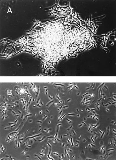
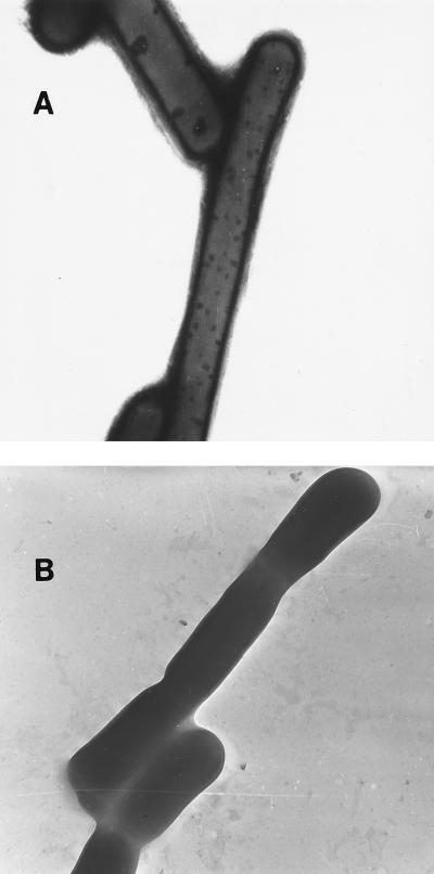
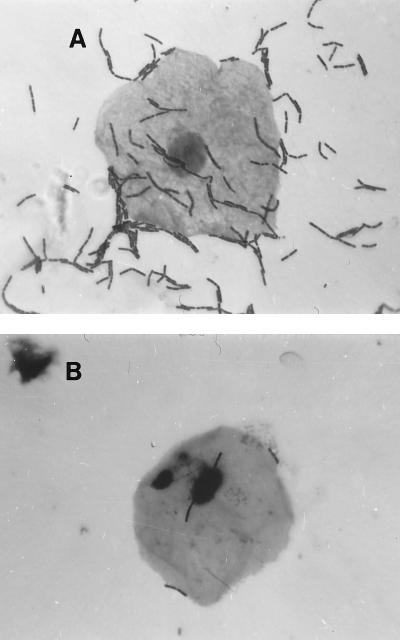
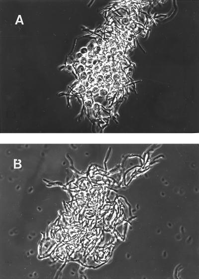
Three strains of Lactobacillus, identified as Lactobacillus acidophilus, Lactobacillus gasseri, and Lactobacillus jensenii, were selected from among 70 isolates from the vaginas of healthy premenopausal women for properties relevant to mucosal colonization or antagonism. All three self-aggregated and adhered to epithelial vaginal cells, displacing well-known vaginal pathogens, such as G. vaginalis, and inhibiting the growth in vitro of Escherichia coli and Streptococcus agalactiae. The surface components involved in self-aggregation appeared to be proteins for L. gasseri and lipoproteins for L. acidophilus and L. jensenii, as judged by susceptibility to treatment with appropriate degrading enzymes. The factors responsible for adherence to epithelial vaginal cells seemed to be glycoproteins (L. acidophilus and L. gasseri) and carbohydrate (L. jensenii). The receptors of the vaginal cells were glycolipids, which presumably were the targets of the competition observed between the lactobacilli and the pathogenic microbes.
Figures
References
-
- Andreu A, Stapleton A E, Fennell C L, Hillier S L, Stamm W E. Hemagglutination, adherence and surface properties of vaginal Lactobacillus species. J Infect Dis. 1995;171:1237–1243. - PubMed
-
- Barrow P A, Brooker P E, Fuller R, Newport M J. The attachment of bacteria to the gastric epithelium of the pig and its importance in the microecology of the intestine. J Appl Bacteriol. 1980;48:147–154. - PubMed
-
- Boris S, Suárez J E, Barbés C. Characterization of the aggregation promoting factor from Lactobacillus gasseri, a vaginal isolate. J Appl Microbiol. 1997;83:413–420. - PubMed
-
- Brooker B E, Fuller R. Adhesion of lactobacilli to the chicken crop epithelium. J Ultrastruct Res. 1975;52:21–31. - PubMed
Publication types
MeSH terms
LinkOut - more resources
Full Text Sources
Other Literature Sources
Medical
