

Cell-contact-stimulated formation of filamentous appendages by Salmonella typhimurium does not depend on the type III secretion system encoded by Salmonella pathogenicity island 1
- PMID: 9573083
- PMCID: PMC108157
- DOI: 10.1128/IAI.66.5.2007-2017.1998
Cell-contact-stimulated formation of filamentous appendages by Salmonella typhimurium does not depend on the type III secretion system encoded by Salmonella pathogenicity island 1
Abstract
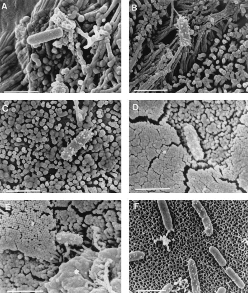
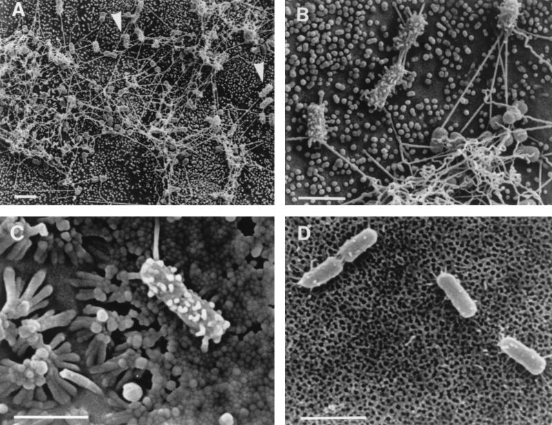
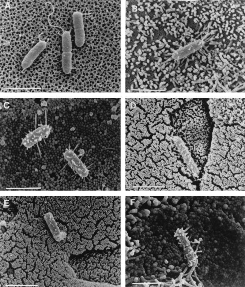
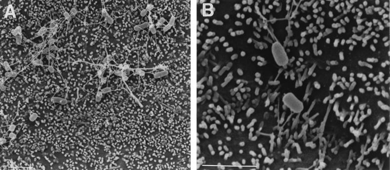
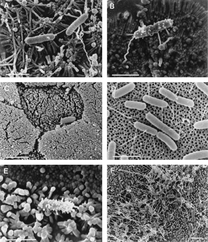
The formation of filamentous appendages on Salmonella typhimurium has been implicated in the triggering of bacterial entry into host cells (C. C. Ginocchio, S. B. Olmsted, C. L. Wells, and J. E. Galán, Cell 76:717-724, 1994). We have examined the roles of cell contact and Salmonella pathogenicity island 1 (SPI1) in appendage formation by comparing the surface morphologies of a panel of S. typhimurium strains adherent to tissue culture inserts, to cultured epithelial cell lines, and to murine intestine. Scanning electron microscopy revealed short filamentous appendages 30 to 50 nm in diameter and up to 300 nm in length on many wild-type S. typhimurium bacteria adhering to both cultured epithelial cells and to murine Peyer's patch follicle-associated epithelia. Wild-type S. typhimurium adhering to cell-free culture inserts lacked these filamentous appendages but sometimes exhibited very short appendages which might represent a rudimentary form of the cell contact-stimulated filamentous appendages. Invasion-deficient S. typhimurium strains carrying mutations in components of SPI1 (invA, invG, sspC, and prgH) exhibited filamentous appendages similar to those on wild-type S. typhimurium when adhering to epithelial cells, demonstrating that formation of these appendages is not itself sufficient to trigger bacterial invasion. When adhering to cell-free culture inserts, an S. typhimurium invG mutant differed from its parent strain in that it lacked even the shorter surface appendages, suggesting that SPI1 may be involved in appendage formation in the absence of epithelia. Our data on S. typhimurium strains in the presence of cells provide compelling evidence that SPI1 is not an absolute requirement for the formation of the described filamentous appendages. However, appendage formation is controlled by PhoP/PhoQ since a PhoP-constitutive mutant very rarely possessed such appendages when adhering to any of the cell types examined.
Figures
Similar articles
-
A PhoP-repressed gene promotes Salmonella typhimurium invasion of epithelial cells.J Bacteriol. 1993 Jul;175(14):4475-84. doi: 10.1128/jb.175.14.4475-4484.1993. J Bacteriol. 1993. PMID: 8392513 Free PMC article.
-
Invasion genes are not required for Salmonella enterica serovar typhimurium to breach the intestinal epithelium: evidence that salmonella pathogenicity island 1 has alternative functions during infection.Infect Immun. 2000 Sep;68(9):5050-5. doi: 10.1128/IAI.68.9.5050-5055.2000. Infect Immun. 2000. PMID: 10948124 Free PMC article.
-
Contact with epithelial cells induces the formation of surface appendages on Salmonella typhimurium.Cell. 1994 Feb 25;76(4):717-24. doi: 10.1016/0092-8674(94)90510-x. Cell. 1994. PMID: 8124710
-
Characterization of effector proteins translocated via the SPI1 type III secretion system of Salmonella typhimurium.Int J Med Microbiol. 2002 Feb;291(6-7):479-85. doi: 10.1078/1438-4221-00156. Int J Med Microbiol. 2002. PMID: 11890547 Review.
-
The Salmonella pathogenicity island-1 type III secretion system.Microbes Infect. 2001 Nov-Dec;3(14-15):1281-91. doi: 10.1016/s1286-4579(01)01488-5. Microbes Infect. 2001. PMID: 11755416 Review.
Cited by
-
Snapshots of usher-mediated protein secretion and ordered pilus assembly.Proc Natl Acad Sci U S A. 2000 Aug 1;97(16):9240-5. doi: 10.1073/pnas.160070497. Proc Natl Acad Sci U S A. 2000. PMID: 10908657 Free PMC article.
-
Intra- and inter-species interactions within biofilms of important foodborne bacterial pathogens.Front Microbiol. 2015 Aug 20;6:841. doi: 10.3389/fmicb.2015.00841. eCollection 2015. Front Microbiol. 2015. PMID: 26347727 Free PMC article. Review.
-
Salmonella enterica serovar Gallinarum requires the Salmonella pathogenicity island 2 type III secretion system but not the Salmonella pathogenicity island 1 type III secretion system for virulence in chickens.Infect Immun. 2001 Sep;69(9):5471-6. doi: 10.1128/IAI.69.9.5471-5476.2001. Infect Immun. 2001. PMID: 11500419 Free PMC article.
-
Polymerization of a single protein of the pathogen Yersinia enterocolitica into needles punctures eukaryotic cells.Proc Natl Acad Sci U S A. 2001 Apr 10;98(8):4669-74. doi: 10.1073/pnas.071065798. Epub 2001 Apr 3. Proc Natl Acad Sci U S A. 2001. PMID: 11287645 Free PMC article.
-
Cytonemes Versus Neutrophil Extracellular Traps in the Fight of Neutrophils with Microbes.Int J Mol Sci. 2020 Jan 16;21(2):586. doi: 10.3390/ijms21020586. Int J Mol Sci. 2020. PMID: 31963289 Free PMC article. Review.
References
-
- Allaoui A, Sansonetti P J, Menard R, Barzu S, Mounier J, Phalipon A, Parsot C. MxiG, a membrane protein required for secretion of Shigella spp. Ipa invasins: involvement in entry into epithelial cells and in intercellular dissemination. Mol Microbiol. 1995;17:461–470. - PubMed
-
- Altmeyer R M, McNern J K, Bossio J C, Rosenshine I, Finlay B B, Galán J E. Cloning and molecular characterisation of a gene involved in Salmonella adherence and invasion of cultured epithelial cells. Mol Microbiol. 1993;7:89–98. - PubMed
-
- Bajaj V, Hwang C, Lee C A. hilA is a novel ompR/toxR family member that activates the expression of Salmonella typhimurium invasion genes. Mol Microbiol. 1995;18:715–727. - PubMed
-
- Bajaj V, Lucas R L, Hwang C, Lee C A. Co-ordinate regulation of Salmonella typhimurium invasion genes by environmental and regulatory factors is mediated by control of hilA expression. Mol Microbiol. 1996;22:703–714. - PubMed
Publication types
MeSH terms
Substances
Grants and funding
LinkOut - more resources
Full Text Sources
