. 1998 May;66(5):2072-7.
doi: 10.1128/IAI.66.5.2072-2077.1998.

Binding of Porphyromonas gingivalis fimbriae to proline-rich glycoproteins in parotid saliva via a domain shared by major salivary components

Affiliations

Binding of Porphyromonas gingivalis fimbriae to proline-rich glycoproteins in parotid saliva via a domain shared by major salivary components

A Amano et al. Infect Immun. 1998 May.

Abstract

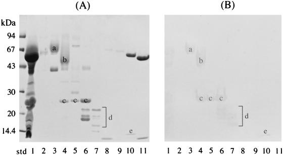
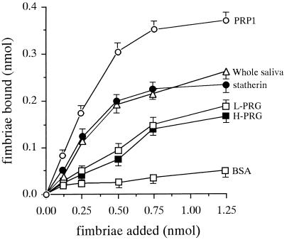
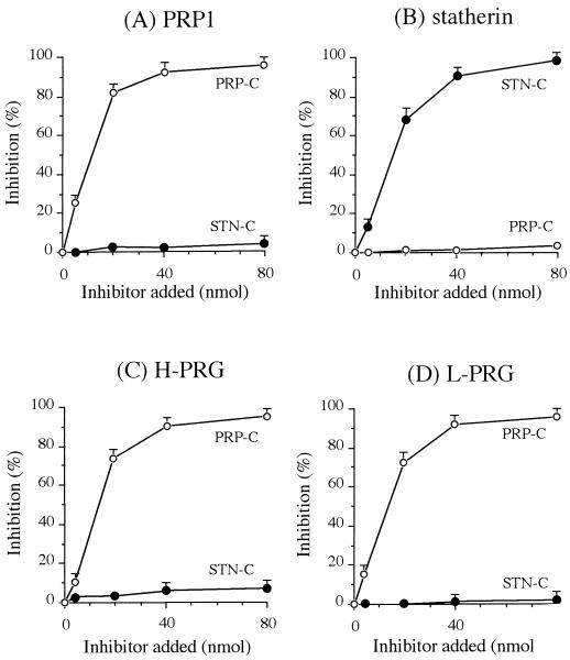
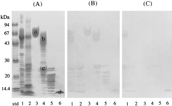
Porphyromonas gingivalis, a putative periodontopathogen, can bind to human saliva through its fimbriae. We previously found that salivary components from the submandibular and sublingual glands bind to P. gingivalis fimbriae and that acidic proline-rich protein (PRP) and statherin function as receptor molecules for fimbriae. In this study, we investigated the fimbria-binding components in parotid saliva. Fractionated human parotid saliva by gel-filtration chromatography was immobilized onto nitrocellulose membranes for the overlay assay following sodium dodecyl sulfate-polyacrylamide gel electrophoresis. The salivary components on the membrane were allowed to interact with fimbriae purified from P. gingivalis ATCC 33277, and the interacted fimbriae were probed with anti-fimbria antibodies. The fimbriae were shown to bind to two forms of proline-rich glycoproteins (PRGs) as well as to acidic PRPs and statherin. Moreover, fimbriae bound to several components of smaller molecular size which appeared to be acidic PRP variants and basic PRPs. Fimbriae bound strongly to the purified PRGs adsorbed onto hydroxyapatite (HAP) beads. In contrast, PRGs in solution failed to inhibit the fimbrial binding to the immobilized PRGs on the HAP beads. These findings suggest that the appearance of binding site(s) of PRGs can be ascribed to their conformational changes. We previously identified the distinct segments within PRP and statherin molecules that are involved in fimbrial binding. The peptides analogous to the binding regions of PRP and statherin (i.e., PRP-C and STN-C) markedly inhibit the binding of fimbriae to PRP and statherin immobilized on the HAP beads, respectively. The PRP-C significantly inhibited the binding of fimbriae to PRG-coated HAP beads as well as to PRP on HAP beads. The peptide did not affect the binding of fimbriae to statherin, whereas the STN-C showed no effect on the fimbrial binding to PRPs or PRGs. In the overlay assay, the PRP-C clearly diminished the interactions between the fimbriae and the various salivary components, including PRPs, the PRGs, and the components with smaller molecular sizes but not statherin. These results strongly suggest that fimbriae bind to salivary components (except statherin) via common peptide segments. It is also suggested that fimbriae bind to saliva through the two distinct binding domains of receptory salivary components: (i) PRGs and PRPs and (ii) statherin.


Figures

FIG. 1
FIG. 1
Binding of parotid saliva components to P. gingivalis fimbriae. (A) SDS-PAGE profiles of parotid saliva and fractionated components. Parotid saliva and the 10 peak fractions were dissolved in SDS sample buffer without heating and were then separated by SDS-PAGE. (B) The salivary components on the nitrocellulose replica were incubated with 5 ml of fimbriae (41 μg/ml; 1 nmol of fimbrillin/ml of KCl buffer). The salivary proteins interacting with fimbriae were probed with anti-fimbria antibodies. It should be noted that PRG and PRPs migrated anomalously with respect to molecular mass in the SDS-PAGE gels. Lanes: std, molecular mass standard; 1, whole parotid saliva; 2 to 11, fractionated parotid salivary components. a and b, novel fimbria-binding proteins found to be PRGs; c, PRP1; d, small-molecular-size components; e, statherin.
FIG. 2
FIG. 2
Binding of P. gingivalis fimbriae to purified PRG-coated HAP beads. HAP beads (3 mg) in a tube were incubated overnight at room temperature with 100 μl of salivary protein solution (100 μg/ml) containing purified PRGs, whole saliva, PRP1, and statherin. 125I-labeled fimbriae were added to a tube containing salivary protein-coated HAP beads and incubated at room temperature for 1 h. The specific binding level was calculated by subtracting the nonspecific binding level, which was obtained by the preincubation of HAP beads with nonlabeled fimbriae (500 μl of 50 nmol/ml) at room temperature for 1 h. All assays were performed in triplicate on three separate occasions. Data are expressed as means ± standard deviations.
FIG. 3
FIG. 3
Effects of H- and L-PRG solutions on the binding of P. gingivalis fimbriae to HAP beads coated with H- and L-PRGs, respectively. Increasing concentrations of H- and L-PRGs in KCl buffer were used as inhibitors for 125I-labeled fimbriae (0.5 nmol). Inhibition studies were performed by the addition of H-PRG in solution to H-PRG-coated HAP beads or of L-PRG to L-PRG-coated HAP beads. All assays were performed in triplicate on three separate occasions. Data are expressed as means ± standard deviations.
FIG. 4
FIG. 4
Effects of the peptides analogous to the carboxyl-terminal segments of PRP1 (PRP-C [PQGPPPQGGRPQGPPQGQSPQ]) and statherin (STN-C [LYPQPYQPQYQQYTF]) on the binding of fimbriae to H- and L-PRG immobilized to HAP beads. Increasing concentrations of the peptides in KCl buffer were used as inhibitors for the binding of 125I-labeled fimbriae (0.5 nmol) to HAP beads coated with salivary proteins: PRP1 (A), statherin (B), H-PRG (C), and L-PRG (D). All assays were performed in triplicate on three separate occasions. Data are expressed as means ± standard deviations.
FIG. 5
FIG. 5
Inhibitory effect of PRP-C on the binding of fimbriae to salivary components. Since the peptide PRP-C (PQGPPPQGGRPQGPPQGQSPQ) was found to be significantly inhibitory for the fimbria-PRG interactions, the overlay assays as shown in Fig. 1 were performed with the addition of PRP-C. (A) SDS-PAGE profiles of salivary components which bound to fimbriae. (B) The overlay assay performed as shown in Fig. 1 without the addition of PRP-C. The replica membrane was incubated with 5 ml of fimbriae (41 μg/ml, 1 nmol/ml of KCl buffer). (C) The overlay assay performed with the simultaneous additions of PRP-C (100 nmol/ml) and fimbriae (1 nmol/ml) in 5 ml of KCl buffer. Lanes: std, molecular mass standard; 1, parotid saliva; 2, submandibular and sublingual saliva; 3, H-PRG; 4, L-PRG–PRP1 fraction; 5, small-molecular-size components that bind to fimbriae; 6, statherin.

Similar articles

Cited by

References

    1. Amano A, Fujiwara T, Nagata H, Kuboniwa M, Sharma A, Sojar H T, Genco R J, Hamada S, Shizukuishi S. Porphyromonas gingivalis fimbriae mediate coaggregation with Streptococcus oralis through specific domains. J Dent Res. 1997;76:852–857. - PubMed
    1. Amano A, Kataoka K, Raj P A, Genco R J, Shizukuishi S. Binding sites of salivary statherin for Porphyromonas gingivalis recombinant fimbrillin. Infect Immun. 1996;64:4249–4254. - PMC - PubMed
    1. Amano A, Sharma A, Lee J-Y, Sojar H T, Raj P A, Genco R J. Structural domains of Porphyromonas gingivalis recombinant fimbrillin that mediate binding to salivary proline-rich protein and statherin. Infect Immun. 1996;64:1631–1637. - PMC - PubMed
    1. Amano A, Sojar H T, Lee J-Y, Sharma A, Levine M J, Genco R J. Salivary receptors for recombinant fimbrillin of Porphyromonas gingivalis. Infect Immun. 1994;62:3372–3380. - PMC - PubMed
    1. Bennick A. Structural and genetic aspects of proline-rich proteins. J Dent Res. 1987;66:457–461. - PubMed

Publication types

LinkOut - more resources