. 1998 May;66(5):2128-34.
doi: 10.1128/IAI.66.5.2128-2134.1998.

Kinetics of infection and effects on placental cell populations in a murine model of Chlamydia psittaci-induced abortion

Affiliations

Kinetics of infection and effects on placental cell populations in a murine model of Chlamydia psittaci-induced abortion

A J Buendía et al. Infect Immun. 1998 May.

Abstract

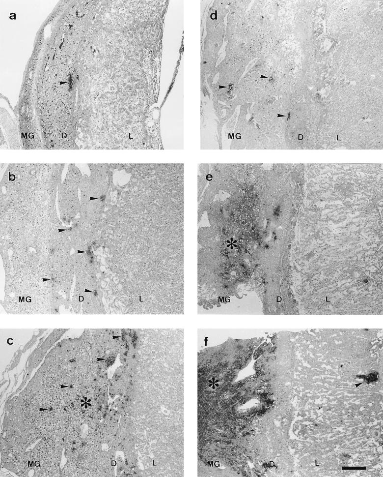
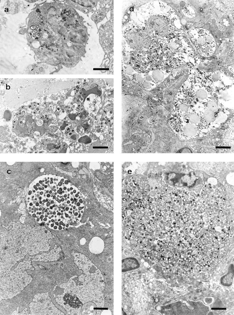
The anatomical progression of chlamydial infection was studied in different areas of the placenta, using a mouse model and two inoculation times: early pregnancy (day 7, group A) and midpregnancy (day 11, group B). The first population cells affected were decidual cells and neutrophils located just at the limits of the maternal and fetal placenta. The following invaded area was the layer of giant cells. Complete colonization of the maternal placenta occurred after day 15 of pregnancy independently of the inoculation time, the metrial gland being the last area to be invaded; numerous granulated metrial gland (GMG) cells were infected. Finally, chlamydial inclusions were observed in labyrinthine trophoblastic cells from day 18 of pregnancy onward. Since no fetal damage was observed, it seems that an indirect mechanism involving the lysis of GMG cells and neutrophil infiltration of the decidua and metrial gland may be the pathogenic mechanism that leads to abortion.


Figures

FIG. 1
FIG. 1
Paraffin wax sections immunostained by the ABC method showing the distribution of positive immunoreaction (arrowheads and asterisks) in the placental areas. Notice that the anatomical progression of infection was increased in group B with regard to group A. (a to c) Group A at 5 days p.i. (small focus in the decidua basalis next to giant-cell layer), 7 days p.i. (small foci disseminated in the decidua basalis), and 9 days p.i. (immunoreaction foci spread through the metrial gland), respectively. (d to f) Group B at 5 days p.i. (small foci disseminated in the decidua basalis and metrial gland), 7 days p.i. (foci forming extensive immunoreaction areas in the decidua basalis and metrial gland), and 9 days p.i. (very extensive immunoreaction area occupying decidua basalis and metrial gland [asterisk] and immunoreaction foci spread through the labyrinth [arrowhead]), respectively. Bar, 266.6 μm for all photographies. MG, metrial gland; D, decidua basalis; L, labyrinth.
FIG. 2
FIG. 2
Electron photomicrographs of the different cell populations affected by chlamydial infection labelled by the immunogold technique. (a) Neutrophil with remains of immunolabelled material within phagolysosomes, day 14 of pregnancy (7 p.i.). Bar, 1.66 μm. (b) Neutrophils with degenerative changes and chromatin condensation containing numerous EBs, day 18 of pregnancy (11 p.i.). Bar, 1.66 μm. (c) Giant cell with a chlamydial inclusion, day 14 of pregnancy (7 p.i.). Bar, 1.98 μm. (d) GMG cell with a young chlamydial inclusion, day 16 of pregnancy (9 p.i.). Bar, 2.64 μm. (e) Decidual cell with a very large chlamydial inclusion, day 18 of pregnancy (11 p.i.). Bar, 1.51 μm.
FIG. 3
FIG. 3
Site of previous attachment at day 2 postabortion. (a) Paraffin wax section immunostained by the ABC technique. Bar, 266.6 μm. (b) Electron photomicrograph labelled by the immunogold technique, showing groups of EBs, amorphous immunolabelled material, and cells with significant degenerative changes. Bar, 1.66 μm.

Similar articles

Cited by

References

    1. Abzug M J, Rotbart H A, Magliato S A, Levin M J. Evolution of the placental barrier to fetal infection by murine enteroviruses. J Infect Dis. 1991;163:1336–1341. - PubMed
    1. Awan A R, Baxi M, Field H J. EHV-1-induced abortion in mice and its relationship to stage of gestation. Res Vet Sci. 1995;59:139–145. - PubMed
    1. Banks J, Eddie B, Schachter J, Meyer K F. Plaque formation by Chlamydia in L cells. Infect Immun. 1970;1:259–262. - PMC - PubMed
    1. Baumgärtner W, Bachmann S. Histological and immunocytochemical characterization of Coxiella burnetii-associated lesions in the murine uterus and placenta. Infect Immun. 1992;60:5232–5241. - PMC - PubMed
    1. Brown M B, Steiner D A. Experimental genital mycoplasmosis: time of infection influences pregnancy outcome. Infect Immun. 1996;64:2315–2321. - PMC - PubMed

Publication types

LinkOut - more resources