. 1998 May;66(5):2170-9.
doi: 10.1128/IAI.66.5.2170-2179.1998.

Generation of neutralizing antipeptide antibodies to the enzymatic domain of Pseudomonas aeruginosa exotoxin A

Affiliations

Generation of neutralizing antipeptide antibodies to the enzymatic domain of Pseudomonas aeruginosa exotoxin A

H S Elzaim et al. Infect Immun. 1998 May.

Abstract

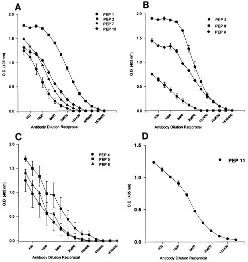
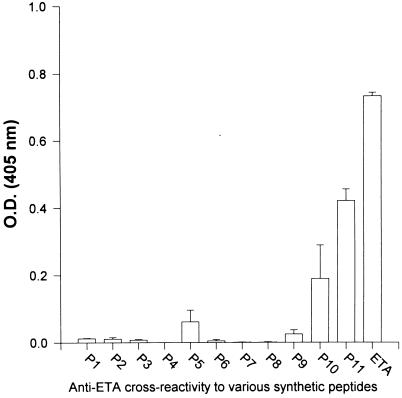
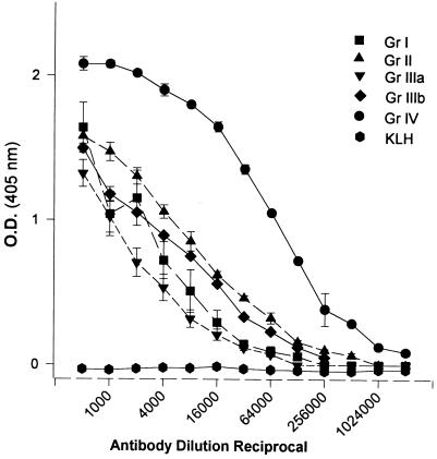
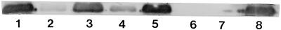
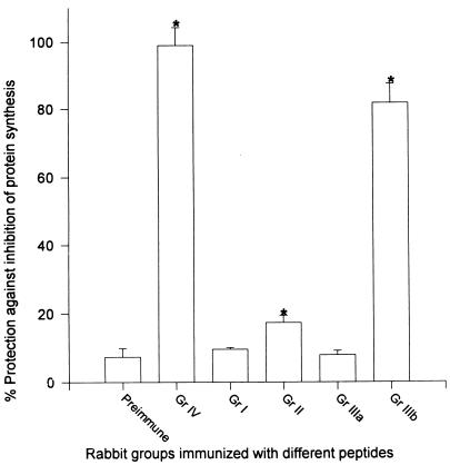
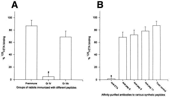
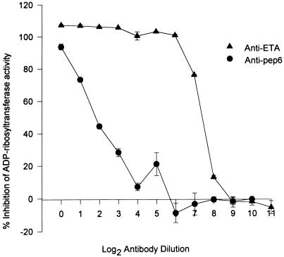
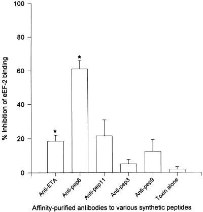
Burn patients suffer a break in the physical barrier (skin), which, when combined with their generalized state of immunodeficiency, creates an open window for opportunistic infections, mainly with Pseudomonas aeruginosa. Infection of the burn wound has always been a major factor in retardation of wound healing, and sepsis remains the leading cause of death in burn patients. Because studies have shown that topical treatment with antiexotoxin A (ETA) antibodies significantly increases survival in rats infected with toxin-producing strains of P. aeruginosa, we examined 11 synthetic peptides encompassing 12 to 45 amino acid (aa) residues, representing what were predicted by computer analysis to be the most hydrophilic and antigenic regions of ETA. These synthetic peptides were injected into rabbits for antibody production. Different groups of rabbits were immunized with a combination of peptides, with each combination representing one of the three distinct domains of ETA. Animals immunized with various peptide combinations produced peptide-specific antibodies that exhibited cross-reactivity to ETA. Two major epitopes were identified on the ETA molecule by experiments with peptide-specific antibodies in enzyme-linked immunosorbent assay and immunoprecipitation. One of these epitopes was located in the translocation domain (II) (aa 297 to 310), while the other was mapped to the last 13 aa residues at the carboxy-terminal end of the enzymatic domain (III) (aa 626 to 638). Of these two regions, the epitope in the enzymatic domain induced a much higher level of neutralizing antibodies that abrogated the cytotoxic activity of ETA in vitro. Antibodies to this epitope blocked the ADP-ribosyltransferase activity of ETA and appeared to interfere with binding of the substrate elongation factor 2 to the enzymatic active site of the ETA molecule. We conclude that polyclonal, as well as monoclonal, antibodies to short peptides, representing small regions of ETA, may have therapeutic potential in passive immunization or topical treatment of burn patients infected with toxin-producing strains of P. aeruginosa.


Figures

FIG. 1
FIG. 1
The 11 synthetic peptides were synthesized, corresponding to different regions within the three structural domains of ETA. The position of the individual peptides in the diagram reflects the location and the overlap between some of them. The sizes of ETA and synthetic peptides were not drawn to scale. The sequences of various synthetic peptides are shown in Table 1.
FIG. 2
FIG. 2
Antibody responses of rabbits immunized with different combinations of peptides (PEP). Serial dilution of serum from individual rabbits was tested by ELISA. The microtiter plates were coated with 1 μg of the corresponding peptide per well. Serum from group I rabbits, immunized with a combination of peptides 1, 2, 7, and 10 (binding domain), was tested against the corresponding peptides (A). Group II rabbits were immunized with a combination of peptides 3, 8, and 9 (translocation domain). The sera from these rabbits were titrated against peptides 3, 8, and 9 (B). Group IIIa rabbits were immunized with a combination of peptides 4, 5, and 6 (enzymatic domain). Their sera were titrated against peptides 4, 5, and 6 (C). Group IIIb rabbits were immunized with peptide 11 (enzymatic domain), and their sera were tested against peptide 11 (D). For each rabbit, serum reactivity to individual peptides was tested in triplicate, and the arithmetic mean ± standard error was plotted for each group of rabbits.
FIG. 3
FIG. 3
Level of peptide (P1, P2, etc.)-cross-reacting antibodies in serum from group IV rabbits (immunized with ETA), as determined by ELISA. Plates were coated with equimolar amounts of individual peptides and ETA (1.5 × 10⁻⁸ M). Serum was tested at a dilution of 1:400. All tests were performed in triplicate, and results were plotted as the mean from two rabbits ± standard error.
FIG. 4
FIG. 4
Titers of sera from different groups (Gr) of rabbits tested against ETA in ELISA plates coated with 100 ng of ETA per well. Group I rabbits were immunized with peptides 1, 2, 7, and 10 (binding domain); group II rabbits were immunized with peptides 3, 8, and 9 (translocation domain); group IIIa rabbits were immunized with peptides 4, 5, and 6 (enzymatic domain); group IIIb rabbits were immunized with peptide 11 only (enzymatic domain); and group IV rabbits were immunized with native ETA. KLH represents a group of two rabbits immunized with KLH (1 mg per rabbit), the carrier protein used in peptide conjugation. Serum samples from each rabbit were tested in triplicate, and the data are plotted as the mean ± standard error for each group of rabbits.
FIG. 5
FIG. 5
Immunoprecipitation of ETA with peptide-specific antibodies. ETA preincubated with serum from group IV (immunized with ETA) rabbits (lane 1), group I (binding domain) rabbits (lane 2), group II (translocation domain) rabbits (lane 3), group IIIa (enzymatic domain) rabbits (lane 4), group IIIb (enzymatic) rabbits (lane 5), preimmune rabbit serum (lane 6), and PBS (lane 7) was precipitated with recombinant protein G, separated on a 12% polyacrylamide gel, transferred to a nitrocellulose membrane, and stained with anti-ETA antibodies (0.25 μg/ml). Lane 8 represents pure ETA (2 μg) directly loaded onto the gel.
FIG. 6
FIG. 6
Protection against ETA-induced inhibition of protein synthesis by antisera raised to different synthetic peptides. Serum samples from different groups (Gr) of rabbits were diluted 1:10 in leucine-deficient EMEM supplemented with [3H]leucine. Two LD50s of ETA (final concentration, 30 ng/ml) were added to the diluted serum, and the mixture was incubated for 1 h at 37°C, before being added to monolayers of 3T3 fibroblasts. After 4 h of incubation at 37°C in the presence of 5% CO2, [3H]leucine incorporated into protein extracted from the cells was measured with a beta counter. Protection was expressed as the percentage of [3H]leucine incorporated in the absence of ETA. Serum samples from each rabbit were tested in triplicate, and the data were plotted as the mean ± standard error for each group. Asterisks indicate statistical significance compared to toxin mixed with preimmune serum (P < 0.05) by one-way analysis of variance.
FIG. 7
FIG. 7
125I-ETA binding to 3T3 fibroblasts in the presence of immune rabbit serum. 125I-ETA was preincubated with a 1:10 dilution of serum from group (Gr) IV rabbits (immunized with ETA) or group IIIb rabbits (immunized with peptide 11) and preimmune serum. The mixture was preincubated for 1 h at 37°C and added to monolayers of 3T3 fibroblasts, which were then incubated for 6 h at 37°C. The level of 125I-ETA remaining after washing was measured with a gamma scintillation counter (A). (B) 125I-ETA was preincubated with affinity-purified antibodies at a concentration 4× the amount necessary to provide 3T3 fibroblasts with 50% protection against inhibition of protein synthesis caused by the same amount of ETA (30 ng/ml). Data were expressed as the percentage of 125I-ETA binding in the absence of antibody. Results represent the mean from two rabbits ± standard error. All experiments were performed in duplicate. An asterisk indicates statistical significance compared to toxin mixed with preimmune serum (A) or PBS (B) (P < 0.05). pep, peptide.
FIG. 8
FIG. 8
Inhibition of ADP-ribosyltransferase activity with immune rabbit serum. ETA (20 ng per reaction mixture) was preincubated for 1 h at 37°C with a 1:5 dilution of preimmune serum or serum from rabbits from group (Gr) I (immunized with peptides [pep] 1, 2, 7, and 10), group II (immunized with peptides 3, 8, and 9), group IIIa (immunized with peptides 4, 5, and 6), group IIIb (immunized with peptide 11), or group IV (immunized with ETA). ETA was then activated, and the reaction mixture was assayed for ADP-ribosylation in the presence of 14C-labeled NAD and eEF-2. The reaction was stopped after 15 min at 25°C with 10% TCA. Samples were then counted with a beta counter (A). (B) ETA (20 ng/ml) was preincubated with 40 μg of affinity-purified peptide-specific antibodies. Inhibition of activity was expressed as the percentage of activity measured in the absence of antibody. Results represent the mean for each group of rabbits ± standard error. Spontaneous ADP-ribosylation of eEF-2 in the absence of ETA was deducted from values obtained in all experiments. Experiments were repeated twice, and each was performed in duplicate. Asterisks indicate statistical significance compared to toxin mixed with preimmune serum (A) or PBS (B) (P < 0.05).
FIG. 9
FIG. 9
Titration of ADP-ribosyltransferase-inhibiting antibodies. Anti-ETA and antipeptide 6 (Anti-pep6) antibodies (2 mg/ml) were serially diluted and incubated with ETA (20 ng per reaction mixture) for 1 h at 37°C. ETA was then activated, and the reaction mixture was incubated for 15 min at 25°C. The reaction was stopped with 10% TCA, and the samples were counted with a beta scintillation counter. Inhibition of activity was expressed as the percentage of activity measured in the absence of antibody. Spontaneous ADP-ribosylation of eEF-2 in the absence of ETA was deducted from the actual values obtained. Results represent the mean ± standard error of two independent experiments, with each experiment performed in duplicate.
FIG. 10
FIG. 10
Inhibition of ETA binding to immobilized eEF-2. ETA (50 ng/ml) was incubated with affinity-purified antibodies (40 μg/well) for 1 h at 37°C. ETA was then activated, and the mixture was added to microtiter plates coated with eEF-2. ETA captured on the plates was then probed with rabbit anti-ETA and HRP-labeled goat anti-rabbit antibodies. The reaction was developed with ABTS, and the OD was determined with an ELISA plate reader. Inhibition of binding was expressed as the percentage of binding measured in the absence of antibody. Bars represent the mean ± standard error from three experiments, with each experiment performed in triplicate. Asterisks represent statistical significance compared to toxin alone (P < 0.05). pep, peptide.

Similar articles

Cited by

References

    1. Allured V S, Collier R J, McKay D B. Structure of exotoxin A of Pseudomonas aeruginosa at 3.0 angstrom resolution. Proc Natl Acad Sci USA. 1986;83:1320–1324. - PMC - PubMed
    1. Beattie B K, Prentice G A, Merrill A R. Investigation into the catalytic role for the tryptophan residues within domain III of Pseudomonas aeruginosa exotoxin A. Biochemistry. 1996;35:15134–15142. - PubMed
    1. Bryan C S, Reynolds X L, Berner E R. Analysis of 1,186 episodes of gram-negative bacteremia in non-university hospitals: the effect of antimicrobial therapy. Rev Infect Dis. 1983;5:629–638. - PubMed
    1. Chaudhary V K, Xu Y H, FitzGerald D, Adhya S, Pastan I. Role of domain II of Pseudomonas exotoxin in the secretion of proteins into the periplasm and medium of Escherichia coli. Proc Natl Acad Sci USA. 1988;85:2939–2943. - PMC - PubMed
    1. Chia J K S, Pollack M, Avigan D, Steinbach S. Functionally distinct monoclonal antibodies reactive with enzymatically active and binding domains of Pseudomonas aeruginosa toxin A. Infect Immun. 1986;52:756–762. - PMC - PubMed

Publication types

MeSH terms

LinkOut - more resources