Changes in murine jejunal morphology evoked by the bacterial superantigen Staphylococcus aureus enterotoxin B are mediated by CD4+ T cells
- PMID: 9573107
- PMCID: PMC108181
- DOI: 10.1128/IAI.66.5.2193-2199.1998
Changes in murine jejunal morphology evoked by the bacterial superantigen Staphylococcus aureus enterotoxin B are mediated by CD4+ T cells
Abstract
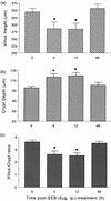
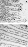
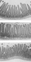
Bacterial superantigens (SAgs) are potent T-cell stimuli that have been implicated in the pathophysiology of autoimmune and inflammatory disease. We used Staphylococcus aureus enterotoxin B (SEB) as a model SAg to assess the effects of SAg exposure on gut form and cellularity. BALB/c, SCID (lacking T cells) and T-cell-reconstituted SCID mice were treated with SEB (5 or 100 microg intraperitoneally), and segments of the mid-jejunum were removed 4, 12, or 48 h later and processed for histochemical or immunocytochemical analysis of gut morphology and major histocompatibility complex class II (MHC II) expression and the enumeration of CD3+ T cells and goblet cells. Control mice received saline only. SEB treatment of BALB/c mice caused a time- and dose-dependent enteropathy that was characterized by reduced villus height, increased crypt depth, and a significant increase in MHC II expression. An increase in the number of CD3+ T cells was observed 48 h after exposure to 100 microg of SEB. Enteric structural alterations were not apparent in SEB-treated SCID mice compared to saline-treated SCID mice. In contrast, SEB challenge of SCID mice reconstituted with a mixed lymphocyte population or purified murine CD4+ T cells resulted in enteric histopathological changes reminiscent of those observed in SEB-treated BALB/c mice. These findings implicate CD4+ T cells in this SEB-induced enteropathy. Our results show that SAg immune activation causes significant changes in jejunal villus-crypt architecture and cellularity that are likely to impact on normal physiological processes. We speculate that the elevated MHC II expression and increased number of T cells could allow for enhanced immune responsiveness to other SAgs or environmental antigens.
Figures


Similar articles
-
CD4+ T cells mediate superantigen-induced abnormalities in murine jejunal ion transport.Am J Physiol. 1998 Jul;275(1):G29-38. doi: 10.1152/ajpgi.1998.275.1.G29. Am J Physiol. 1998. PMID: 9655681
-
Nitric oxide participates in the recovery of normal jejunal epithelial ion transport following exposure to the superantigen, Staphylococcus aureus enterotoxin B.J Immunol. 1999 Oct 15;163(8):4519-26. J Immunol. 1999. PMID: 10510395
-
Colonic bacterial superantigens evoke an inflammatory response and exaggerate disease in mice recovering from colitis.Gastroenterology. 2003 Dec;125(6):1785-95. doi: 10.1053/j.gastro.2003.09.020. Gastroenterology. 2003. PMID: 14724831
-
MAIT cells launch a rapid, robust and distinct hyperinflammatory response to bacterial superantigens and quickly acquire an anergic phenotype that impedes their cognate antimicrobial function: Defining a novel mechanism of superantigen-induced immunopathology and immunosuppression.PLoS Biol. 2017 Jun 20;15(6):e2001930. doi: 10.1371/journal.pbio.2001930. eCollection 2017 Jun. PLoS Biol. 2017. PMID: 28632753 Free PMC article.
-
Therapeutic down-modulators of staphylococcal superantigen-induced inflammation and toxic shock.Toxins (Basel). 2010 Aug;2(8):1963-83. doi: 10.3390/toxins2081963. Epub 2010 Jul 29. Toxins (Basel). 2010. PMID: 22069668 Free PMC article. Review.
Cited by
-
Superantigen immune stimulation evokes epithelial monocyte chemoattractant protein 1 and RANTES production.Infect Immun. 1999 Nov;67(11):6198-202. doi: 10.1128/IAI.67.11.6198-6202.1999. Infect Immun. 1999. PMID: 10531290 Free PMC article.
-
Staphylococcal enterotoxins.Toxins (Basel). 2010 Aug;2(8):2177-97. doi: 10.3390/toxins2082177. Epub 2010 Aug 18. Toxins (Basel). 2010. PMID: 22069679 Free PMC article. Review.
-
A Case of Staphylococcus aureus Enterocolitis: A Rare Entity.Gastroenterol Hepatol (N Y). 2010 Feb;6(2):117-9. Gastroenterol Hepatol (N Y). 2010. PMID: 20567554 Free PMC article. No abstract available.
-
Identification of a transcytosis epitope on staphylococcal enterotoxins.Infect Immun. 2002 Apr;70(4):2178-86. doi: 10.1128/IAI.70.4.2178-2186.2002. Infect Immun. 2002. PMID: 11895985 Free PMC article.
-
Local and global consequences of flow on bacterial quorum sensing.Nat Microbiol. 2016 Jan 11;1:15005. doi: 10.1038/nmicrobiol.2015.5. Nat Microbiol. 2016. PMID: 27571752 Free PMC article.
References
-
- Aisenberg J, Ebert E C, Mayer L. T cell activation in human intestinal mucosa: the role of superantigens. Gastroenterology. 1993;105:1421–1430. - PubMed
-
- Ansell J D, Bancroft G J. The biology of the SCID mutation. Immunol Today. 1989;10:322–325. - PubMed
-
- Blackman M A, Woodland D L. In vivo effects of superantigens. Life Sci. 1995;57:1717–1735. - PubMed
-
- Braegger C P, MacDonald T T. Immune mechanisms in chronic inflammatory bowel disease. Ann Allergy. 1994;72:135–141. - PubMed
Publication types
MeSH terms
Substances
LinkOut - more resources
Full Text Sources
Other Literature Sources
Research Materials

