. 1998 May;66(5):2300-9.
doi: 10.1128/IAI.66.5.2300-2309.1998.

Essential role of gamma interferon in survival of colon ascendens stent peritonitis, a novel murine model of abdominal sepsis

Affiliations
Free PMC article

Essential role of gamma interferon in survival of colon ascendens stent peritonitis, a novel murine model of abdominal sepsis

N Zantl et al. Infect Immun. 1998 May.
Free PMC article

Abstract

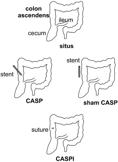
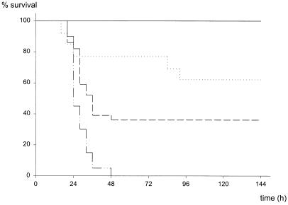
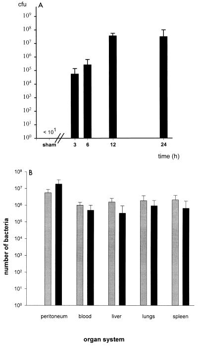
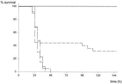
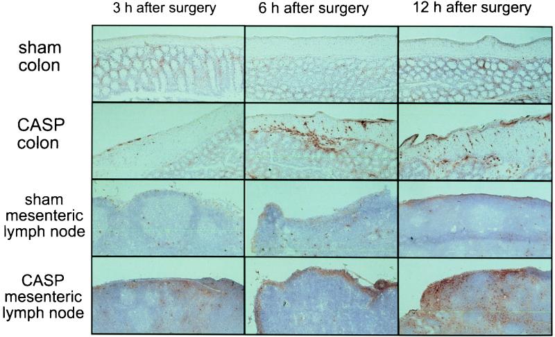
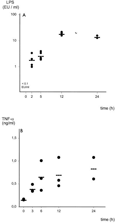
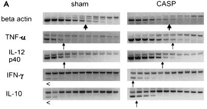
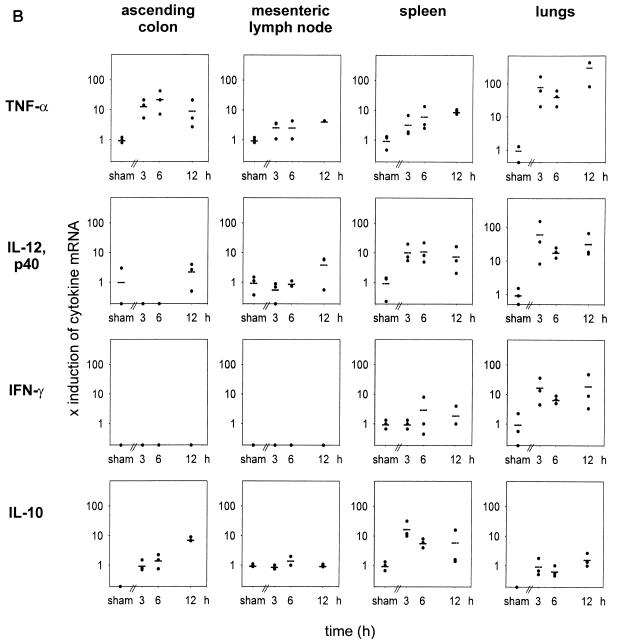
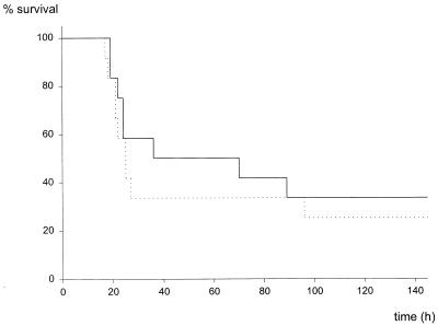
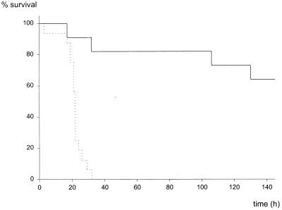
Despite considerable progress, peritonitis and sepsis remain life-threatening conditions. To improve the understanding of the pathophysiology encountered in sepsis, a new standardized and highly reproducible murine model of abdominal sepsis termed colon ascendens stent peritonitis (CASP) was developed. In CASP, a stent is inserted into the ascending colon, which generates a septic focus. CASP employing a stent of 14-gauge diameter (14G stent) results in a mortality of 100% within 18 to 48 h after surgery. By inserting stents of small diameters, mortality can be exactly controlled. Thus, CASP surgery with insertion of a 22G or 18G stent (22G or 18G CASP surgery) results in 38 or 68% mortality, respectively. 14G CASP surgery leads to a rapid invasion of bacteria into the peritoneum and the blood. As a consequence, endotoxemia occurs, inflammatory cells are recruited, and a systemic inflammatory response syndrome develops. Interestingly, the most pronounced upregulation of inflammatory cytokines (gamma interferon [IFN-gamma], tumor necrosis factor alpha [TNF-alpha] and interleukin-12) is observed in spleen and lungs. CASP surgery followed by stent removal at specific time intervals revealed that all animals survived if intervention was performed after 3 h, whereas removal of the septic focus after 9 h did not prevent death, suggesting induction of autonomous mechanisms of a lethal inflammatory response syndrome. 18G CASP surgery in IFN-gamma receptor-deficient (IFNgammaR-/-) mice revealed an essential role of IFN-gamma in survival of sepsis, whereas TNF receptor p55-deficient (TNFRp55-/-) mice did not show altered survival rates. In summary, this study describes a novel animal model that closely mimics human sepsis and appears to be highly suitable for the study of the pathophysiology of abdominal sepsis. Importantly, this model demonstrates a protective role of IFN-gamma in survival of bacterial sepsis.


Figures

FIG. 1
FIG. 1
Schematic principle of CASP, sham CASP, and CASPI surgery. In CASP surgery, a stent of a defined diameter is introduced into the ascending colon by puncture and fixed with a suture to the colonic wall. Insertion of the stent allows transmigration of colonic flora from the gut into the peritoneal cavity. In sham CASP surgery, the stent is fixed to the colonic wall with a suture without puncturing the colonic lumen. In CASPI, the stent is surgically removed and the defect of the colonic wall is closed by a sinking suture as described by Lembert (34a). In sham CASPI, a defect measuring the diameter of the stent is cut into the wall of the ascending colon and immediately closed by surgery as in CASPI.
FIG. 2
FIG. 2
Mortality after CASP surgery using different stent diameters. After insertion of stents of different diameters (22, 18, or 14 gauge), survival of mice was closely monitored. Mice scoring more than 8 points on the sepsis score (from 0 [healthy] to 12 [comatose]) were sacrificed. ——, sham CASP (n = 9); ·····, 22G CASP (n = 13); — —, 18-G CASP (n = 44); —···, 14-G CASP (n = 20).
FIG. 3
FIG. 3
(A) Bacterial counts in the peritoneal cavity. The peritoneal cavity was flushed with sterile saline solution 3, 6, 12, and 24 h after CASP surgery. Titrated serial dilutions of the lavage fluid were plated on selective agar plates, and bacterial CFU were calculated. After sham CASP surgery, no bacteria were detected in peritoneal lavage fluid. (B) Bacteria counts and species in different organ systems. Mice were sacrificed 12 h after 14G CASP surgery or sham surgery. Peritoneal lavage was performed, blood was collected, and lungs, liver, and spleen were harvested under sterile conditions as described in the text. Organs were homogenized in 4 ml of sterile PBS buffer. Then 10 μl-aliquots of 10-fold dilutions of blood, peritoneal lavage fluid, or organ suspensions were plated on blood agar and MacConkey plates and incubated under both aerobic and anaerobic conditions. Bacterial counts are given as number of bacteria per whole organ, entire volume of peritoneal lavage, or milliliter of blood (sample means ± standard deviation, n = 7 mice for peritoneal lavage and peripheral blood, n = 3 mice for liver, lungs, and spleen). All specimens of sham-operated control mice were sterile (data not shown). ░⃞, gram-negative bacteria; ▪, gram-positive bacteria.
FIG. 4
FIG. 4
Survival after stent removal (CASPI). CASP surgery employing a 14G stent was performed at a time point defined as 0 h. After 3, 5, or 9 h, the stent was surgically removed and the colonic wall was closed. Sham CASPI was performed as described in Materials and Methods and the legend to Fig. 1. ——, sham CASPI (n = 11); ·····, 14G CASPI, stent removal after 3 h (n = 13); ——, 14G CASPI, stent removal after 5 h (n = 25); —···, 14G CASPI, stent removal after 9 h (n = 13); —, 14G CASP (n = 20).
FIG. 5
FIG. 5
Infiltration of tissues with inflammatory cells. Recruitment of inflammatory cells was examined by immunohistochemical staining of colon and mesenteric lymph node cryosections of sham- or 14G CASP-operated mice, which were sacrificed 3, 6, or 12 h after surgery. Ascending colon and mesenteric lymph nodes were removed at given points in time, sectioned, and stained with biotinylated CD11b (Mac-1α) monoclonal antibody followed by streptavidin-peroxidase. The stain was reacted with 3-aminoethylcarbazole (see Materials and Methods). CD11b-positive cells (granulocytes and monocytes/macrophages) are labeled in red.
FIG. 6
FIG. 6
(A) LPS amounts in the bloodstream of mice that underwent CASP surgery. Plasma amounts of LPS (endotoxin) were determined in 14G CASP-operated mice at given points in time by conducting a KQCL test (see Materials and Methods). LPS amounts in healthy mice (0 h) and sham-operated mice (data not shown) were below the detection limit of the LPS assay of 0.005 EU/ml. As early as 2 h after induction of sepsis, substantial amounts of LPS could be detected. (B) Kinetics of TNF serum amounts after CASP surgery. TNF-α content in the plasma of mice after 14G CASP surgery was determined by ELISA.
FIG. 7
FIG. 7
Detection of cytokine mRNA in organs after CASP surgery. (A) Internal competitive semiquantitative cytokine RT-PCR from spleens harvested 6 h after 14G CASP or sham surgery. mRNA was extracted from spleens of mice after sham or 14G CASP surgery, and cDNA was transcribed. cDNAs were serially diluted, and the content of cDNA was estimated by internal competitive semiquantitative RT-PCR with β-actin-specific primers in the presence of known amounts of β-actin control fragment. TNF-α, IL-12p40, IFN-γ, and IL-10 cDNA amounts were determined by PCR amplification of serial dilutions from equilibrated cDNA amounts in the presence of the relevant PCR primers (Table 1) and the corresponding control fragment. The upper band represents the amplified control fragment of a known constant concentration, whereas the lower band shows the signal obtained after amplification of each titrated cytokine cDNA. The arrows indicate the concentrations of equal amounts of control fragment and cytokine cDNA. Upregulation of mRNAs for TNF-α, IL-12p40, and, to a smaller extent, IFN-γ can be observed. (B) Kinetics of the induction of cytokine mRNA transcription in colon, mesenteric lymph nodes, spleen, and lungs after CASP and sham surgery. Upregulation of TNF-α, IL-12p40, IFN-γ, and IL-10 was analyzed 3, 6, and 12 h after 14G CASP or sham surgery in various organs of mice; ascending colon, mesenteric lymph nodes, spleen, and lungs were removed, RNA was extracted, and cDNA was prepared. Semiquantitative PCR was performed as described for panel A. Induction of cytokine mRNA was calculated as fold induction over basal levels as determined after sham surgery. For explanation of calculations, see text.
FIG. 7
FIG. 7
Detection of cytokine mRNA in organs after CASP surgery. (A) Internal competitive semiquantitative cytokine RT-PCR from spleens harvested 6 h after 14G CASP or sham surgery. mRNA was extracted from spleens of mice after sham or 14G CASP surgery, and cDNA was transcribed. cDNAs were serially diluted, and the content of cDNA was estimated by internal competitive semiquantitative RT-PCR with β-actin-specific primers in the presence of known amounts of β-actin control fragment. TNF-α, IL-12p40, IFN-γ, and IL-10 cDNA amounts were determined by PCR amplification of serial dilutions from equilibrated cDNA amounts in the presence of the relevant PCR primers (Table 1) and the corresponding control fragment. The upper band represents the amplified control fragment of a known constant concentration, whereas the lower band shows the signal obtained after amplification of each titrated cytokine cDNA. The arrows indicate the concentrations of equal amounts of control fragment and cytokine cDNA. Upregulation of mRNAs for TNF-α, IL-12p40, and, to a smaller extent, IFN-γ can be observed. (B) Kinetics of the induction of cytokine mRNA transcription in colon, mesenteric lymph nodes, spleen, and lungs after CASP and sham surgery. Upregulation of TNF-α, IL-12p40, IFN-γ, and IL-10 was analyzed 3, 6, and 12 h after 14G CASP or sham surgery in various organs of mice; ascending colon, mesenteric lymph nodes, spleen, and lungs were removed, RNA was extracted, and cDNA was prepared. Semiquantitative PCR was performed as described for panel A. Induction of cytokine mRNA was calculated as fold induction over basal levels as determined after sham surgery. For explanation of calculations, see text.
FIG. 8
FIG. 8
Mortality of TNFRp55−/− mice after CASP surgery. TNFRp55+/+ (——) and TNFRp55−/− (·····) (41) animals (C57BL/6 background) were subjected to 18G CASP surgery. Survival was monitored. Four of 12 mice in the control group and 3 of 12 TNFRp55−/− mice survived 18G CASP.
FIG. 9
FIG. 9
Mortality of IFNγR−/− mice after CASP surgery. IFNγR−/− (32) (·····) and IFNγR+/+ (——) control littermates (C57BL/6 × 129/Sv background) were analyzed after 18G CASP surgery. Seven of 11 IFNγR+/+ mice survived, whereas all of the 15 mice with an inactivated receptor for IFN-γ died.

Similar articles

Cited by

References

    1. Abraham E, Glauser M P, Butler T, Garbino J, Gelmont D, Laterre P F, Kudsk K, Bruining H A, Otto C, Tobin E, Zwingelstein C, Lesslauer W, Leighton A. p55 tumor necrosis factor receptor fusion protein in the treatment of patients with severe sepsis and septic shock. A randomized controlled multicenter trial. Ro 45-2081 Study Group. JAMA. 1997;277:1531–1538. - PubMed
    1. Anonymous. American College of Chest Physicians/Society of Critical Care Medicine Consensus Conference: definitions for sepsis and organ failure and guidelines for the use of innovative therapies in sepsis. Crit Care Med. 1992;20:864–874. - PubMed
    1. Bach E A, Aguet M, Schreiber R D. The IFN gamma receptor: a paradigm for cytokine receptor signaling. Annu Rev Immunol. 1997;15:563–591. - PubMed
    1. Bagby G J, Plessala K J, Wilson L A, Thompson J J, Nelson S. Divergent efficacy of antibody to tumor necrosis factor-alpha in intravascular and peritonitis models of sepsis. J Infect Dis. 1991;163:83–88. - PubMed
    1. Baker C C, Chaudry I H, Gaines H O, Baue A E. Evaluation of factors affecting mortality rate after sepsis in a murine cecal ligation and puncture model. Surgery. 1983;94:331–335. - PubMed

Publication types

Substances