. 1998 May;66(5):2310-8.
doi: 10.1128/IAI.66.5.2310-2318.1998.

Differential early interactions between Salmonella enterica serovar Typhi and two other pathogenic Salmonella serovars with intestinal epithelial cells

Affiliations

Differential early interactions between Salmonella enterica serovar Typhi and two other pathogenic Salmonella serovars with intestinal epithelial cells

D L Weinstein et al. Infect Immun. 1998 May.

Abstract

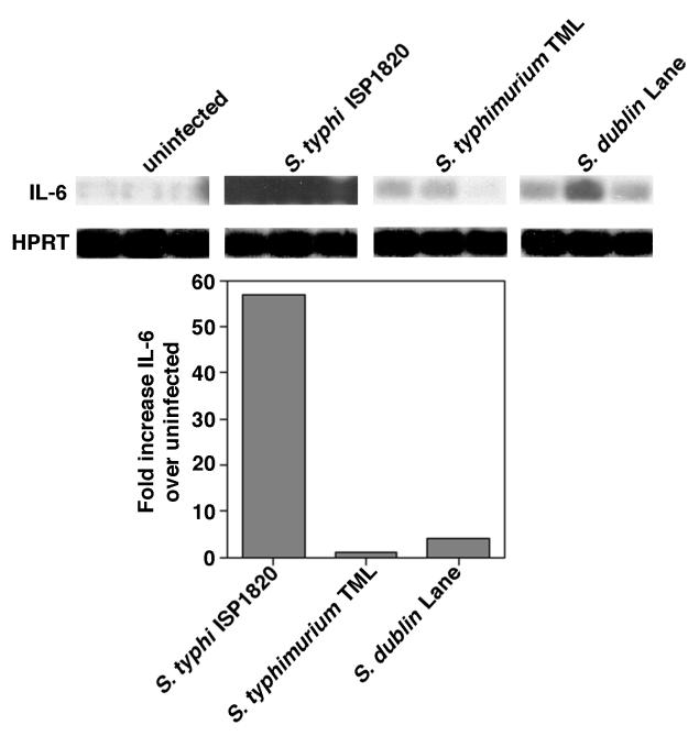
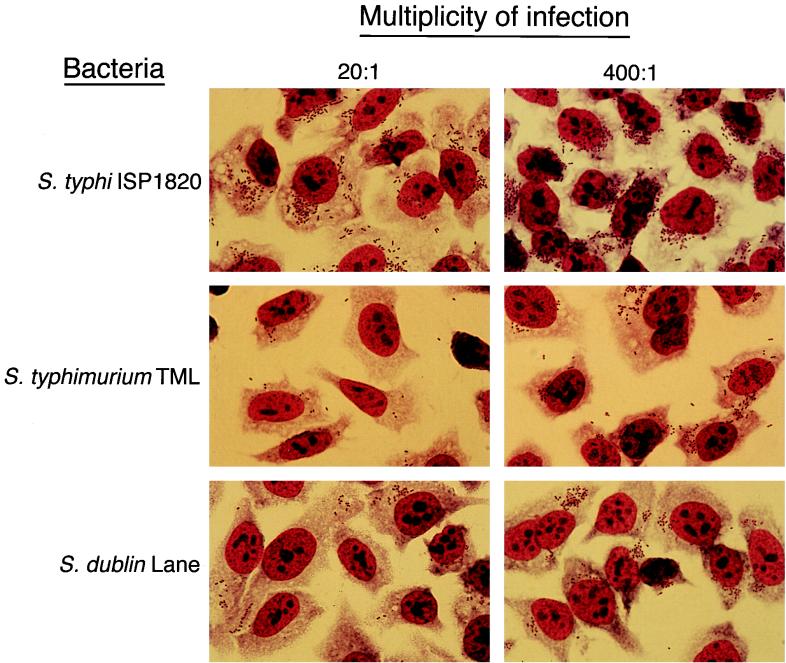
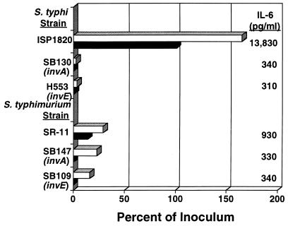
Salmonella enterica serovar Typhi (hereafter referred to as S. typhi) is a host-restricted pathogen that adheres to and invades the distal ileum and subsequently disseminates to cause typhoid fever in humans. However, S. typhi appears to be avirulent in small animals. In contrast, other pathogenic salmonellae, such as S. enterica serovars Typhimurium and Dublin (S. typhimurium and S. dublin, respectively), typically cause localized gastroenteritis in humans but have been used as models for typhoid fever because these organisms cause a disease in susceptible rodents that resembles human typhoid. In vivo, S. typhi has been demonstrated to attach to and invade murine M cells but is rapidly cleared from the Peyer's patches without destruction of the M cells. In contrast, invasion of M cells by S. typhimurium is accompanied by destruction of these M cells and subsequently sloughing of the epithelium. These data have furthered our view that the early steps in the pathogenesis of typhoidal and nontyphoidal Salmonella serovars are distinct. To extend this concept, we have utilized an in vitro model to evaluate three parameters of initial host-pathogen interactions: adherence of three Salmonella serovars to human and murine small intestinal epithelial cell (IEC) lines, the capacity of these salmonellae to invade IECs, and the ability of the bacteria to induce interleukin-6 (IL-6) in these cell lines as a measure of host cell activation and the host acute-phase response. The results demonstrate that S. typhi adheres to and invades human small IECs better than either S. typhimurium or S. dublin. Interestingly, invA and invE null mutants of S. typhi are able neither to adhere to nor to invade IECs, unlike S. typhimurium invA and invE mutants, which adhere to but cannot invade IECs. S. typhi also induces significantly greater quantities of IL-6 in human small IEC lines than either of the other two Salmonella serovars. These findings suggest that differential host cytokine responses to bacterial pathogens may play an important role in the pathological sequelae that follow infection. Importantly, S. typhimurium did not induce IL-6 in murine IECs. Since S. typhimurium infection in mice is often used as a model of typhoid fever, these findings suggest that, at least in this case, the mouse model does not reflect the human disease. Taken together, our studies indicate that (i) marked differences occur in the initial steps of S. typhi, S. typhimurium, and S. dublin pathogenesis, and (ii) conclusions about S. typhi pathogenesis that have been drawn from the mouse model of typhoid fever should be interpreted conservatively.


Figures

FIG. 1
FIG. 1
Cytokine gene expression by Int407 stimulated with S. typhi ISP1820, S. typhimurium TML, and S. dublin Lane. RNA was prepared from 2.6 × 10⁶ uninfected Int407 cells or cells stimulated with S. typhi ISP1820, S. typhimurium TML, or S. dublin Lane at MOIs of approximately 20 bacteria per cell in a standard invasion assay. cDNA was prepared from 1 μg of each mRNA sample, and RT-PCR was performed with primers specific for human IL-6 and the housekeeping gene HPRT. Blots were hybridized with enhanced chemiluminescence-labeled oligonucleotide probes specific for the corresponding genes. Autoradiographs were scanned, and the fold increase of the IL-6-specific cytokine gene was calculated by comparison of the signals elicited by bacteria with those elicited by medium alone. Each lane of the autoradiograph represents the RT-PCR product from a single well. These results are representative of three separate experiments.
FIG. 2
FIG. 2
The effects of extending the incubation periods postinfection on invasion and IL-6 induction of Salmonella-infected Int407 cells. Monolayers of Int407 cells (2.5 × 10⁵ cells per well of a 24-well tissue culture plate) were infected with an MOI of 20 S. typhi ISP1820 (squares), S. typhimurium TML (diamonds), or S. dublin Lane (circles) bacteria per cell and incubated for 90 min to allow the bacteria to adhere and invade. After removal of the extracellular bacteria by washing, the cultures were further incubated in the presence of gentamicin for an additional 90, 120, 180, or 240 min. At the end of the culture period, the supernatants were removed, and the concentration of IL-6 in each supernatant was determined by bioassay. The Int407 cells were lysed, and the number of bacteria surviving gentamicin treatment was determined. These results are representative of two independent experiments.
FIG. 3
FIG. 3
Effects of increasing the MOI on invasion and IL-6 induction of Salmonella-infected Int407 cells. Monolayers of Int407 cells (2.5 × 10⁵ cells per well of a 24-well tissue culture plate) were infected with various doses of S. typhi ISP1820 (squares), S. typhimurium TML (circles), or S. dublin Lane (diamonds) and incubated for 90 min to allow the bacteria to adhere and invade. After removal of the extracellular bacteria by washing, the cultures were further incubated in the presence of gentamicin for an additional 90 min. At the end of the culture period, the supernatants were removed, and the concentration of IL-6 in each supernatant was determined by bioassay. The Int407 cells were lysed, and the number of bacteria surviving gentamicin treatment was determined. These results are representative of four independent experiments. The values plotted for S. typhi ISP1820 are taken from the work of Weinstein et al. (47).
FIG. 4
FIG. 4
Microscopic comparison of the adherence and invasion profiles of S. typhi ISP1820-, S. typhimurium TML-, and S. dublin Lane-infected Int407 cells. Semiconfluent monolayers of Int407 cells (8 × 10⁵ cells per well of a six-well tissue culture plate) were overlaid with medium (uninfected control) or infected at a bacterium-to-cell ratio of 20:1 or 400:1 with S. typhi ISP1820, S. typhimurium TML, or S. dublin Lane and incubated for 90 min to allow the bacteria to adhere and invade. Cultures were washed, fixed, and then stained with Fisher Leukostat staining reagents. The monolayers were photographed under the oil immersion objective (×100 magnification) of an Olympus BX50 microscope. These results are representative of three independent experiments.
FIG. 5
FIG. 5
Effects of invA and invE mutations on the adherence, invasion, and IL-6 induction of S. typhi ISP1820 and S. typhimurium TML on Int407 cells. Monolayers of Int407 cells (2.5 × 10⁵ cells per well of a 24-well tissue culture plate) were overlaid with medium (uninfected control) or infected at a bacterium-to-cell ratio of 20:1 with S. typhi ISP1820, S. typhi SB130 (ISP1820 invA), S. typhi H553 (ISP1820 invE), S. typhimurium SR-11, S. typhimurium SB147 (SR-11 invA), or S. typhimurium SB109 (SR-11 invE) and incubated for 90 min to allow the bacteria to adhere and invade. Cultures were washed and incubated in the presence of gentamicin for an additional 90 min to eliminate extracellular bacteria. At the end of the culture period, the supernatants were removed, and the concentration of IL-6 in each supernatant was determined by B9 bioassay. The IECs were lysed, and the number of viable intracellular bacteria was determined. These results are representative of two independent experiments. Top and bottom bars for each strain represent percent cell association and percent invasion, respectively.

References

    1. Agace W W, Hedges S R, Ceska M, Svanborg C. Interleukin-8 and the neutrophil response to mucosal gram-negative infection. J Clin Invest. 1993;92:780–785. - PMC - PubMed
    1. Carter P B, Collins F M. Growth of typhoid and paratyphoid bacilli in intravenously infected mice. Infect Immun. 1974;10:816–822. - PMC - PubMed
    1. Clark M A, Jepson M A, Simmons N L, Hirst B H. Preferential interaction of Salmonella typhimurium with mouse Peyer’s patch M cells. Res Microbiol. 1994;145:543–552. - PubMed
    1. Collins F M, Carter P B. Growth of salmonellae in orally infected germfree mice. Infect Immun. 1978;21:41–47. - PMC - PubMed
    1. Crabtree J E, Wyatt J I, Trejdosiewicz L K, Peichl P, Nichols P H, Ramsay N, Primrose J N, Lindley I J D. Interleukin-8 expression in Helicobacter pylori infected, normal, and neoplastic gastroduodenal mucosa. J Clin Pathol. 1994;47:61–66. - PMC - PubMed

Publication types

LinkOut - more resources