

Identification of an Na+-dependent malonate transporter of Malonomonas rubra and its dependence on two separate genes
- PMID: 9573154
- PMCID: PMC107221
- DOI: 10.1128/JB.180.10.2689-2693.1998
Identification of an Na+-dependent malonate transporter of Malonomonas rubra and its dependence on two separate genes
Abstract
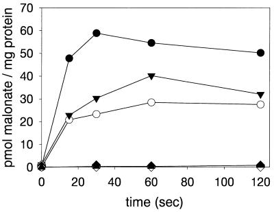
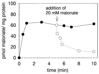
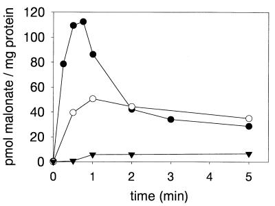
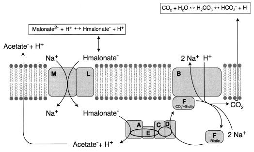
Two membrane proteins encoded by the malonate fermentation gene cluster of Malonomonas rubra, MadL and MadM, have been synthesized in Escherichia coli. MadL and MadM were shown to function together as a malonate transport system, whereas each protein alone was unable to catalyze malonate transport. Malonate transport by MadLM is Na+ dependent, and imposition of a DeltapNa+ markedly enhanced the rate of malonate uptake. The kinetics of malonate uptake into E. coli BL21(DE3) cells synthesizing MadLM at different pH values indicated that Hmalonate- is the transported malonate species. The stimulation of malonate uptake by Na+ ions showed Michaelis-Menten kinetics, and a Km for Na+ of 1.2 mM was determined. These results suggest that MadLM is an electroneutral Na+/Hmalonate- symporter and that it is dependent on two separate genes.
Figures
Similar articles
-
Sequence of a gene cluster from Malonomonas rubra encoding components of the malonate decarboxylase Na+ pump and evidence for their function.Eur J Biochem. 1997 Apr 1;245(1):103-15. doi: 10.1111/j.1432-1033.1997.00103.x. Eur J Biochem. 1997. PMID: 9128730
-
The fermenting bacterium Malonomonas rubra is phylogenetically related to sulfur-reducing bacteria and contains a c-type cytochrome similar to those of sulfur and sulfate reducers.Syst Appl Microbiol. 1998 Aug;21(3):340-5. doi: 10.1016/S0723-2020(98)80042-8. Syst Appl Microbiol. 1998. PMID: 9841124
-
Enzymic and genetic basis for bacterial growth on malonate.Mol Microbiol. 1997 Jul;25(1):3-10. doi: 10.1046/j.1365-2958.1997.4611824.x. Mol Microbiol. 1997. PMID: 11902724 Review.
-
The biotin protein MadF of the malonate decarboxylase from Malonomonas rubra.Arch Microbiol. 1998 Nov;170(6):464-8. doi: 10.1007/s002030050668. Arch Microbiol. 1998. PMID: 9799291
-
Malonate metabolism: biochemistry, molecular biology, physiology, and industrial application.J Biochem Mol Biol. 2002 Sep 30;35(5):443-51. doi: 10.5483/bmbrep.2002.35.5.443. J Biochem Mol Biol. 2002. PMID: 12359084 Review.
Cited by
-
Genome Sequence of Clinical Strain Pseudomonas aeruginosa NRD619.Microbiol Resour Announc. 2020 Oct 29;9(44):e01013-20. doi: 10.1128/MRA.01013-20. Microbiol Resour Announc. 2020. PMID: 33122414 Free PMC article.
-
Biocatalytic synthesis of 2-fluoro-3-hydroxypropionic acid.Front Bioeng Biotechnol. 2022 Aug 17;10:969012. doi: 10.3389/fbioe.2022.969012. eCollection 2022. Front Bioeng Biotechnol. 2022. PMID: 36061447 Free PMC article.
-
Engineering a Novel Metabolic Pathway for Improving Cellular Malonyl-CoA Levels in Escherichia coli.Mol Biotechnol. 2023 Sep;65(9):1508-1517. doi: 10.1007/s12033-022-00635-5. Epub 2023 Jan 19. Mol Biotechnol. 2023. PMID: 36658293
-
Engineering controllable alteration of malonyl-CoA levels to enhance polyketide production.Nat Chem Biol. 2025 Aug;21(8):1214-1225. doi: 10.1038/s41589-025-01911-6. Epub 2025 Jun 11. Nat Chem Biol. 2025. PMID: 40500421 Free PMC article.
-
Engineered Fluorine Metabolism and Fluoropolymer Production in Living Cells.Angew Chem Int Ed Engl. 2017 Oct 23;56(44):13637-13640. doi: 10.1002/anie.201706696. Epub 2017 Sep 26. Angew Chem Int Ed Engl. 2017. PMID: 28861937 Free PMC article.
References
-
- Berg M, Hilbi H, Dimroth P. Sequence of a gene cluster from Malonomonas rubra encoding components of the malonate decarboxylase Na+ pump and evidence for their function. Eur J Biochem. 1997;245:103–115. - PubMed
-
- Bott M. Anaerobic citrate metabolism and its regulation in enterobacteria. Arch Microbiol. 1997;167:78–88. - PubMed
-
- Bradford M M. A rapid and sensitive method for the quantitation of microgram quantities of protein utilizing the principle of protein-dye binding. Anal Biochem. 1976;72:248–254. - PubMed
-
- Cornish-Bowden A. Fundamentals of enzyme kinetics. London, United Kingdom: Portland Press; 1995.
-
- Dehning I, Schink B. Malonomonas rubra gen. nov. sp. nov., a microaerotolerant anaerobic bacterium growing by decarboxylation of malonate. Arch Microbiol. 1989;151:427–433.
MeSH terms
Substances
LinkOut - more resources
Full Text Sources
Other Literature Sources
