Comparative Study
. 1998 May;180(9):2298-305.
doi: 10.1128/JB.180.9.2298-2305.1998.

Nucleotide sequence and spatiotemporal expression of the Vibrio cholerae vieSAB genes during infection

Affiliations
Comparative Study

Nucleotide sequence and spatiotemporal expression of the Vibrio cholerae vieSAB genes during infection

S H Lee et al. J Bacteriol. 1998 May.

Abstract

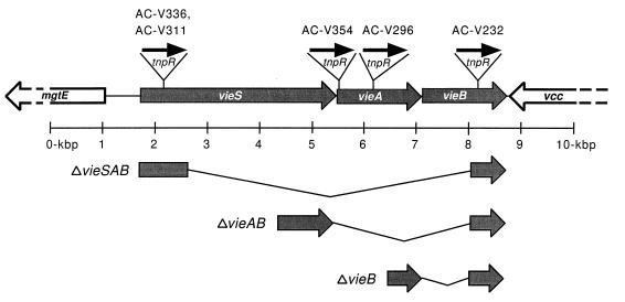
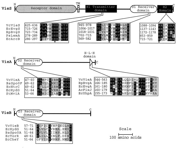
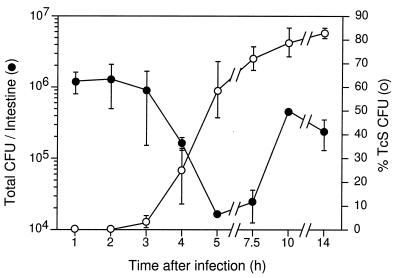
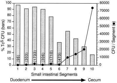
The iviVII gene of Vibrio cholerae was previously identified by a screen for genes induced during intestinal infection. In the present study, nucleotide sequence analysis revealed that iviVII is a 1,659-bp open reading frame, herein designated vieB, that is predicted to be last in a tricistronic operon (vieSAB). The deduced amino acid sequence of VieS exhibited similarity to the sensor kinase component, and those of VieA and VieB were similar to the response regulator components, respectively, of the two-component signal transduction family. Analysis of transcriptional fusions to a site-specific DNA recombinase reporter, tnpR, revealed that vieS and vieA are transcribed during in vitro growth in a vieAB-independent and vieA-dependent manner, respectively. In contrast, transcription of vieB occurred exclusively during infection and was not dependent upon VieB. We conclude that the vieSAB genes are differentially regulated, at least during laboratory growth. Use of a V. cholerae strain harboring a vieB::tnpR transcriptional fusion allowed the kinetics and location of vieB expression within the intestine to be determined. We found that vieB transcription is induced shortly after infection of the proximal and mid-small intestine.


Figures

FIG. 1
FIG. 1
Genetic organization of the V. cholerae vie genes. The vieSAB genes are flanked by a partially sequenced, divergently transcribed ORF designated mgtE and a partially sequenced, convergently transcribed ORF designated vcc. The sites of the transcriptional fusions in strains AC-V311, AC-V336, AC-V354, AC-V296, and AC-V232 are shown at the top. The flanking sequences used to construct in-frame deletion mutations are shown at the bottom along with the corresponding deleted regions, designated by the thin angled lines.
FIG. 2
FIG. 2
Schematic representations of VieS, VieA, and VieB, and multiple alignments of conserved domains characteristic of two-component signal transduction proteins. Identical residues are indicated by black squares, and functionally similar residues are indicated by gray squares. Conserved phosphorylated histidine and aspartate residues are indicated below by an asterisk. Protein domains in the alignments are numbered and are designated by the abbreviated species name joined to the gene name. Abbreviations: Vc, V. cholerae; Ec, E. coli; Bp, B. pertussis; Ps, Pseudomonas syringae; Bs, Bacillus subtilis; St, Salmonella typhimurium.
FIG. 3
FIG. 3
Kinetics of transcriptional induction of vieB during infection. V. cholerae AC-V232 (vieB::tnpR-lacZY) was used to intragastrically inoculate infant CD-1 mice. At the postinoculation times indicated on the x axis, the small intestines were removed and homogenized. Total CFU per intestine, shown on the left axis, was determined by plating serial dilutions on agar medium. The percent Tcs CFU per intestine, shown on the right axis, was determined by replica plating. Each time point was investigated in triplicate, i.e., three animals were used per time point, and the means and standard deviations are shown.
FIG. 4
FIG. 4
Localization of the intestinal segment where vieB transcription is induced. V. cholerae AC-V232 (vieB::tnpR-lacZY) was used to intragastrically inoculate three infant CD-1 mice. At 3.5 h postinoculation, the small intestines and cecum were removed and dissected into 10 segments of equal length. The total CFU, shown on the right axis, and percent Tcs CFU (bars), shown on the left axis, were determined for each segment. The total CFU for segments 1 to 6 are shown in parentheses above each data point. The data in this figure are from one animal and are representative of the results found with the other two animals.

Similar articles

Cited by

References

    1. Altschul S F, Gish W, Miller W, Myers E W, Lipman D J. Basic local alignment search tool. J Mol Biol. 1990;215:403–410. - PubMed
    1. Arico B, Miller J F, Roy C, Stibitz S, Monack D, Falkow S, Gross R, Rappuoli R. Sequences required for expression of Bordetella pertussis virulence factors share homology with prokaryotic signal transduction proteins. Proc Natl Acad Sci USA. 1989;86:6671–6675. - PMC - PubMed
    1. Attridge S R, Voss E, Manning P A. The role of toxin-coregulated pili in the pathogenesis of Vibrio cholerae O1 El Tor. Microb Pathog. 1993;15:421–431. - PubMed
    1. Camilli A, Beattie D, Mekalanos J. Use of genetic recombination as a reporter of gene expression. Proc Natl Acad Sci USA. 1994;91:2634–2638. - PMC - PubMed
    1. Camilli A, Mekalanos J J. Use of recombinase gene fusions to identify Vibrio cholerae genes induced during infection. Mol Microbiol. 1995;18:671–683. - PMC - PubMed

Publication types

Substances

Associated data

LinkOut - more resources