. 1998 May;180(9):2312-20.
doi: 10.1128/JB.180.9.2312-2320.1998.

Cloning, sequence analysis, and characterization of the genes involved in isoprimeverose metabolism in Lactobacillus pentosus

Affiliations

Cloning, sequence analysis, and characterization of the genes involved in isoprimeverose metabolism in Lactobacillus pentosus

S Chaillou et al. J Bacteriol. 1998 May.

Abstract

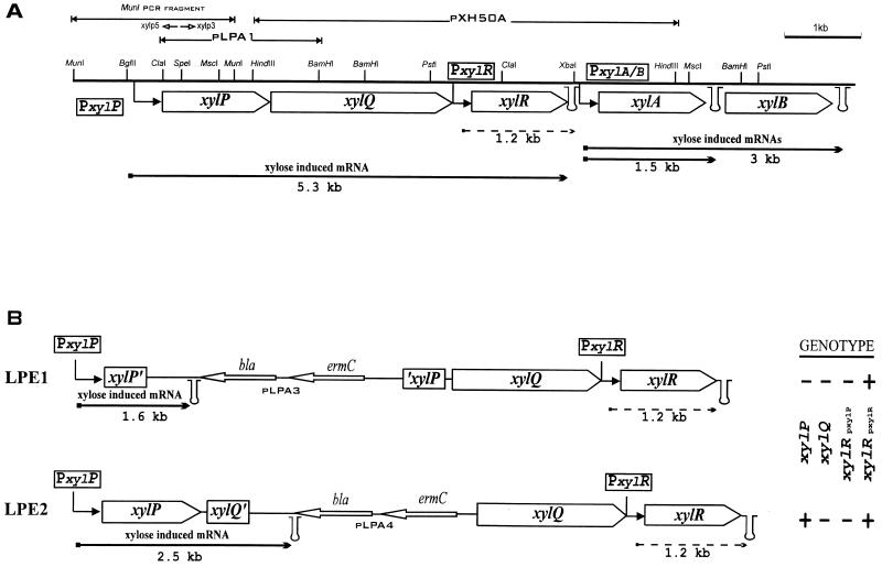
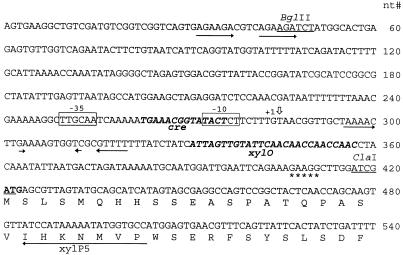
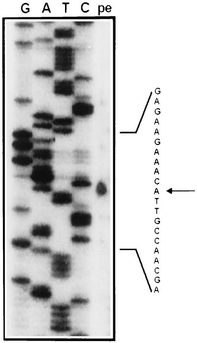
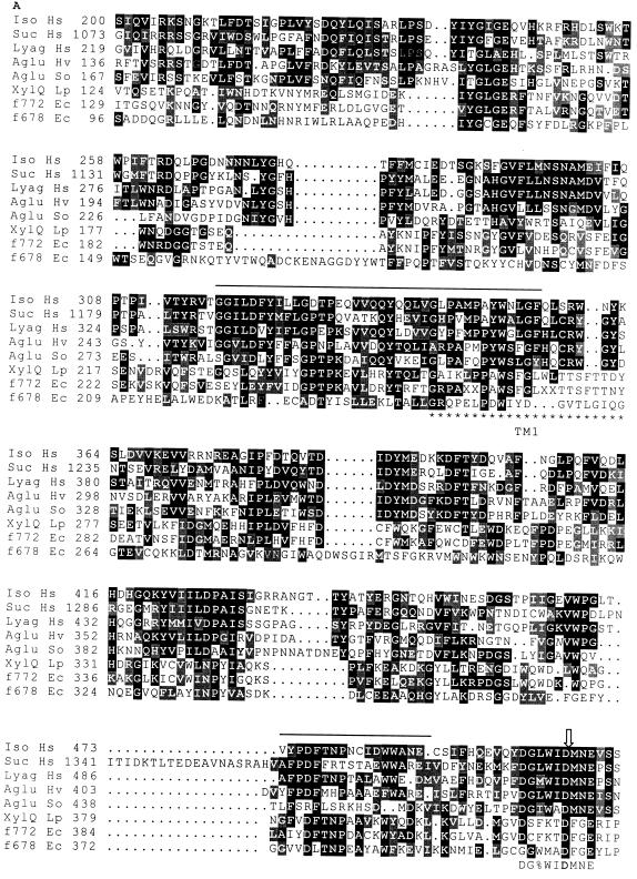
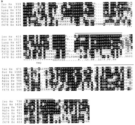
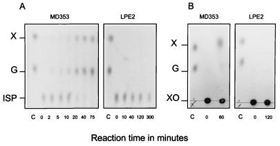
Two genes, xylP and xylQ, from the xylose regulon of Lactobacillus pentosus were cloned and sequenced. Together with the repressor gene of the regulon, xylR, the xylPQ genes form an operon which is inducible by xylose and which is transcribed from a promoter located 145 bp upstream of xylP. A putative xylR binding site (xylO) and a cre-like element, mediating CcpA-dependent catabolite repression, were found in the promoter region. L. pentosus mutants in which both xylP and xylQ (LPE1) or only xylQ (LPE2) was inactivated retained the ability to ferment xylose but were impaired in their ability to ferment isoprimeverose (alpha-D-xylopyranosyl-(1,6)-D-glucopyranose). Disruption of xylQ resulted specifically in the loss of a membrane-associated alpha-xylosidase activity when LPE1 or LPE2 cells were grown on xylose. In the membrane fraction of wild-type bacteria, alpha-xylosidase could catalyze the hydrolysis of isoprimeverose and p-nitrophenyl-alpha-D-xylopyranoside with apparent Km and Vmax values of 0.2 mM and 446 nmol/min/mg of protein, and 1.3 mM and 54 nmol/min/mg of protein, respectively. The enzyme could also hydrolyze the alpha-xylosidic linkage in xyloglucan oligosaccharides, but neither methyl-alpha-D-xylopyranoside nor alpha-glucosides were substrates. Glucose repressed the synthesis of alpha-xylosidase fivefold, and 80% of this repression was released in an L. pentosus delta ccpA mutant. The alpha-xylosidase gene was also expressed in the absence of xylose when xylR was disrupted.


Figures

FIG. 1
FIG. 1
(A) Physical map and organization of the L. pentosus MD353 xylose regulon. The upper part shows the xylP xylQ cloning strategy. (B) Chromosomal situations of LPE1 and LPE2 integrants after integration of pLPA3 and pLPA4 plasmids. Their respective genotypes are indicated on the right (plus and minus signs indicate the presence and absence, respectively, of transcription of the genes). In both panels, arrows with right angles indicate the xylPQ and xylAB (xylose-inducible) promoters and the xylR (constitutive) promoters, and stem-loop structures indicate the putative transcriptional terminators. The sizes of the wild-type bacterial, LPE1, and LPE2 transcripts from the different promoters are given below each arrow (arrows with solid and dashed lines represent xylose-inducible and constitutive expression, respectively). The primers used for the inverse PCR, xylp5 and xylp3, are indicated by short open arrows below the MunI fragment. bla and ermC, genes for ampicillin resistance and erythromycin resistance, respectively.
FIG. 2
FIG. 2
Nucleotide (nt) sequence of the promoter-regulatory region of the xylPQR operon. Open boxes, −10 and −35 elements of the promoter; boldface italic letters, putative regulatory elements (cre and xylO); stars, potential ribosome binding site; open vertical arrow, +1 transcription start; horizontal arrow, sequence complementary to the primer xylp5 used in the primer extension experiment. The beginning of the deduced amino acid sequence of the xylP gene is depicted below the nucleotide sequence. Direct repeated or inverted repeated sequences in the promoter region are underlined with arrows.
FIG. 3
FIG. 3
Primer extension analysis of xylose-induced RNA transcript from L. pentosus MD353. The MunI PCR-amplified fragment was used as the template for the sequencing reaction. Lane pe contains the primer extension reaction with the kinase-reacted xylp5 primer. G, guanine; A, adenosine; T, thymine; C, cytosine. The start of the transcript is indicated by an arrow.
FIG. 4
FIG. 4
Alignments of similar regions of primary structure of isomaltase (Iso Hs) and sucrase (Suc Hs) from the human pro-sucrase-isomaltase complex (Swiss-Prot database accession no. P14410), the human lysosomal α-glucosidase (Lyag Hs; accession no. P10253), the Schwanniomyces occidentalis glucoamylase (Aglu So; P22861), the barley putative α-glucosidase (Aglu Hv) (34), and the three prokaryotic polypeptides L. pentosus XylQ (XylQ Lp) and the E. coli hypothetical proteins f772 (f772 Ec; P31434) and f678 (f678 Ec; P32138). Identical amino acids are shown in white letters on a solid ground, and similar amino acids are shown in white letters on a shaded ground. Stretches of amino acids revealed by the BLAST research are overlined. The PROSITE signatures (PDOC00120) of family 31 of glycosyl hydrolases are shown below the alignment, as follows: %, any one of L, I, V, or M; $, either F or Y; #, either S or T; !, either S or A. The open arrow points to the aspartic acid residue involved in the active site of the human lysosomal α-glucosidase. Stars under the sequence mark the putative transmembrane helices of XylQ. Numbers in front of the lines are amino acid positions of the proteins.
FIG. 4
FIG. 4
Alignments of similar regions of primary structure of isomaltase (Iso Hs) and sucrase (Suc Hs) from the human pro-sucrase-isomaltase complex (Swiss-Prot database accession no. P14410), the human lysosomal α-glucosidase (Lyag Hs; accession no. P10253), the Schwanniomyces occidentalis glucoamylase (Aglu So; P22861), the barley putative α-glucosidase (Aglu Hv) (34), and the three prokaryotic polypeptides L. pentosus XylQ (XylQ Lp) and the E. coli hypothetical proteins f772 (f772 Ec; P31434) and f678 (f678 Ec; P32138). Identical amino acids are shown in white letters on a solid ground, and similar amino acids are shown in white letters on a shaded ground. Stretches of amino acids revealed by the BLAST research are overlined. The PROSITE signatures (PDOC00120) of family 31 of glycosyl hydrolases are shown below the alignment, as follows: %, any one of L, I, V, or M; $, either F or Y; #, either S or T; !, either S or A. The open arrow points to the aspartic acid residue involved in the active site of the human lysosomal α-glucosidase. Stars under the sequence mark the putative transmembrane helices of XylQ. Numbers in front of the lines are amino acid positions of the proteins.
FIG. 5
FIG. 5
TLC analysis of the products of hydrolysis of isoprimeverose (A) or xyloglucan oligosaccharides (B) by membrane fractions of wild-type L. pentosus MD353 and the LPE2 mutant. Lane C, xylose and glucose control (5 nmol of each). X, xylose; G, glucose; ISP, isoprimeverose; XO, xyloglucan oligosaccharides. The reactions were performed in 50 μl of KPED buffer (pH 6.5) at 37°C. The reaction mixtures contained 10 μg of membrane proteins and either 60 nmol of isoprimeverose or 1.8 μmol of xylogucan oligosaccharides. At each time point, 1/10 of the reaction mixture was loaded on the TLC plates.

Similar articles

Cited by

References

    1. Altschul S F, Gish W, Miller W, Myers E W, Lipman D J. Basic local alignment search tool. J Mol Biol. 1990;215:403–410. - PubMed
    1. Bergmeyer H U. Methods of enzymatic analysis. VI. Deerfield Beach, Fla: VCH Publishers; 1985.
    1. Burland V D, Plunkett III G, Daniels D L, Blattner F R. DNA sequence and analysis of 136 kilobases of the Escherichia coli genome: organisational symmetry around the origin of replication. Genomics. 1993;16:551–561. - PubMed
    1. Callens M, Kerstens-Hilderson H, van Opstal O, de Bruine C K. Catalytic properties of d-xylose isomerase from Streptomyces violaceoruber. Enzyme Microb Technol. 1986;8:696–700.
    1. Daeschel M A, Andersson R E, Fleming H P. Microbial ecology of fermenting plant materials. FEMS Microbiol Rev. 1987;46:357–367.

Publication types

MeSH terms

Associated data

LinkOut - more resources