. 1998 May;180(9):2321-9.
doi: 10.1128/JB.180.9.2321-2329.1998.

The 102-kilobase unstable region of Yersinia pestis comprises a high-pathogenicity island linked to a pigmentation segment which undergoes internal rearrangement

Affiliations

The 102-kilobase unstable region of Yersinia pestis comprises a high-pathogenicity island linked to a pigmentation segment which undergoes internal rearrangement

C Buchrieser et al. J Bacteriol. 1998 May.

Abstract

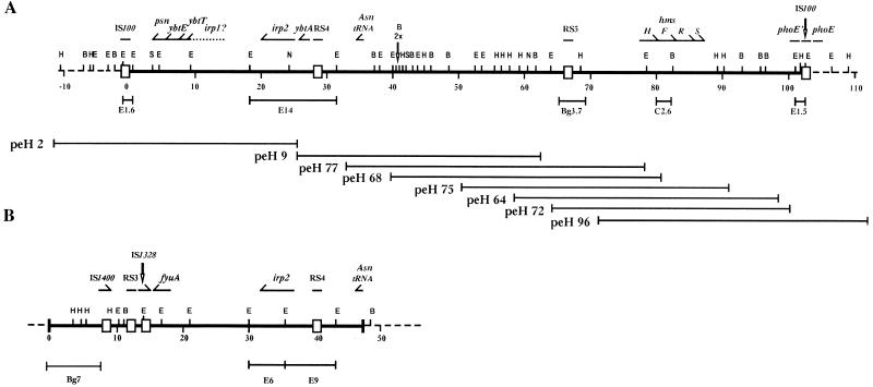
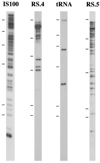
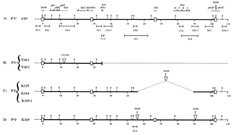
Several pathogenicity islands have recently been identified in different bacterial species, including a high-pathogenicity island (HPI) in Yersinia enterocolitica 1B. In Y. pestis, a 102-kb chromosomal fragment (pgm locus) that carries genes involved in iron acquisition and colony pigmentation can be deleted en bloc. In this study, characterization and mapping of the 102-kb region of Y. pestis 6/69 were performed to determine if this unstable region is a pathogenicity island. We found that the 102-kb region of Y. pestis is composed of two clearly distinct regions: an approximately 35-kb iron acquisition segment, which is an HPI per se, linked to an approximately 68-kb pigmentation segment. This linkage was preserved in all of the Y. pestis strains studied. However, several nonpigmented Y. pestis strains harboring an irp2 gene have been previously identified, suggesting that the pigmentation segment is independently mobile. Comparison of the physical map of the 102-kb region of these strains with that of strain 6/69 and complementation experiments were carried out to determine the genetic basis of this phenomenon. We demonstrate that several different mechanisms involving mutations and various-size deletions are responsible for the nonpigmented phenotype in the nine strains studied. However, no deletion corresponded exactly to the pigmentation segment. The 102-kb region of Y. pestis is an evolutionarily stable linkage of an HPI with a pigmentation segment in a region of the chromosome prone to rearrangement in vitro.


Figures

FIG. 1
FIG. 1
Restriction maps of the 102-kb region of Y. pestis 6/69 (A) and the HPI of Y. enterocolitica Ye8081 (B). Plain thick lines, the 102-kb region; broken lines, regions outside the 102-kb region. Restriction sites: E, EcoRI; H, HindIII; B, BamHI; S, SpeI; N, NotI. Horizontal bars below the restriction maps represent the different probes cited in the text. The values below the map indicate the scale in kilobases. Horizontal arrows and bars above the restriction map correspond to the identified genes. peH, cosmid clones used to span the entire 102-kb region. RS, repeated sequences; 2×, two very close BamHI restriction sites.
FIG. 2
FIG. 2
Hybridization profiles of four different repeated sequences present in the 102-kb region of strain 6/69. The EcoRI-digested genomic DNA of strain 6/69 was hybridized with the E1.6 probe from Y. pestis 6/69 for IS100 (Fig. 1A), the SCI1 probe from Y. enterocolitica Ye8081 for RS.4 (5), a 50-bp oligonucleotide probe from Y. enterocolitica Ye8081 for the asn tRNA (5), and the Bg3.7 probe from Y. pestis 6/69 for RS.5. The tick marks indicate the following molecular sizes, top to bottom: 23.1, 9.4, 6.5, 4.3, 2.3, and 2.0 kb.
FIG. 3
FIG. 3
Comparison of the 102-kb region EcoRI restriction map of strain 6/69 (A) with those of strains from Turkey (B) and Kenya (C and D). Plain thick lines, the 102-kb region; broken lines, regions outside the 102-kb region; dotted lines, deleted regions; E, EcoRI restriction sites; triangles, DNA sequences that were absent from the 6/69 102-kb region. Horizontal bars below the restriction maps represent the different probes cited in the text. The values below the map indicate the scale in kilobases. Horizontal arrows and bars above the restriction map correspond to the identified genes. RS, repeated sequences; P+I+, Pgm+ Irp2+; PI+, Pgm Irp2+.
FIG. 4
FIG. 4
Comparison of the 102-kb region EcoRI restriction map of strain 6/69 (A) with those of strains from Hamburg (B), Ho Chi Minh City (C), and Zaire (D). Plain thick lines, the 102-kb region; broken lines, regions outside the 102-kb region; E, EcoRI restriction sites; triangles, DNA sequences that were absent from the 6/69 102-kb region. Horizontal bars below the restriction maps represent the different probes cited in the text and used to analyze the strains. The values below the map indicate the scale in kilobases. Horizontal arrows and bars above the restriction map correspond to the identified genes. RS, repeated sequences.

Similar articles

Cited by

References

    1. Bearden S W, Fetherston J D, Perry R D. Genetic organization of the yersiniabactin biosynthetic region and construction of avirulent mutants in Yersinia pestis. Infect Immun. 1997;65:1659–1668. - PMC - PubMed
    1. Birnboim H C, Doly J. A rapid alkaline extraction procedure for screening recombinant plasmid DNA. Nucleic Acids Res. 1979;7:1513–1523. - PMC - PubMed
    1. Blum G, Ott M, Lischewski A, Ritter A, Imrich H, Tschäpe H, Hacker J. Excision of large DNA regions termed pathogenicity islands from tRNA-specific loci in the chromosome of an Escherichia coli wild-type pathogen. Infect Immun. 1994;62:606–614. - PMC - PubMed
    1. Buchrieser C, Buchrieser O, Kristl A, Kaspar C W. Clamped homogeneous electric fields (CHEF) gel-electrophoresis of DNA restriction fragments for comparing genomic variations among strains of Yersinia enterocolitica and Yersinia spp. Zentralbl Bakteriol. 1994;281:457–470. - PubMed
    1. Carniel E, Guilvout I, Prentice M. Characterization of a large chromosomal “high-pathogenicity island” in biotype 1B Yersinia enterocolitica. J Bacteriol. 1996;178:6743–6751. - PMC - PubMed

Publication types

MeSH terms